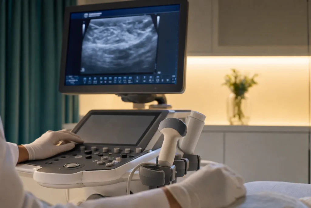
Fibroadenoma
Finding a lump in your breast is unsettling, and it is natural to fear the worst. But a great many breast lumps are not cancer. A fibroadenoma is one of the most common of these benign (not cancer) lumps, especially in younger women. This guide explains what a fibroadenoma is, why it forms, how doctors confirm it, and the calm, evidence-based choices you have for managing it.
What a fibroadenoma is
A fibroadenoma is a benign (not cancer) lump in the breast. The word looks complicated, but it simply describes what the lump is made of: fibro- means fibrous (firm, supportive) tissue, and -adenoma means a growth made of gland tissue. So a fibroadenoma is a small, solid ball of the normal building blocks of the breast — the milk-making gland tissue and the connective tissue that holds it together — that has grown together into a distinct lump.
Fibroadenomas are the most common benign breast lumps. They are most often found in women in their 20s and 30s, though they can appear at any age after periods begin, and they become uncommon after menopause. They are not contagious, they are not your fault, and having one does not mean you did anything wrong.
Most importantly: a typical (simple) fibroadenoma is not cancer and does not turn into cancer. Doctors still confirm the diagnosis carefully, because any new breast lump deserves proper checking, but the great majority turn out to be harmless. Understanding that early can take a lot of the fear out of the weeks of testing ahead.
Types and subtypes
Not all fibroadenomas are the same. Knowing the type helps you understand what to expect and which treatment makes sense.
- Simple fibroadenoma. By far the most common type. Under the microscope the cells look uniform and orderly. A simple fibroadenoma does not increase your future breast cancer risk.
- Complex fibroadenoma. When the pathologist (the doctor who examines tissue) looks at the cells, there are some extra changes, such as small cysts or calcium deposits. These tend to be a little larger and are more common in women over about 35. A complex fibroadenoma may carry a very slightly higher risk of breast cancer in the future compared with women who have no breast lumps — but the increase is small, and your team will explain what it means for you.
- Giant fibroadenoma. A fibroadenoma larger than about 5 centimetres (roughly 2 inches). Removal is more often advised simply because of its size and the way it can distort the breast.
- Juvenile fibroadenoma. A type seen in teenage girls. It can grow quickly during the teenage years but is still benign.
One related lump is worth a brief mention. A phyllodes tumour can feel like a fibroadenoma but behaves differently and is usually removed. This is one reason doctors sometimes recommend taking a sample or removing a lump that grows — to be sure of exactly what it is.
Causes and risk factors
The honest answer is that doctors do not know exactly what causes a fibroadenoma. It is not caused by injury, by diet, or by anything you did. The strongest clue points to hormones, particularly oestrogen (the main female sex hormone).
Fibroadenomas seem to be sensitive to oestrogen. That fits with what is observed in real life: they tend to appear during the reproductive years, can grow during pregnancy or while taking hormones (such as some hormone therapy), and often shrink after menopause when oestrogen levels fall. This is a normal response of breast tissue to its natural hormonal environment, not a sign that something has gone wrong.
A few patterns make a fibroadenoma more likely, though none of them are things to feel anxious about:
- Being a younger woman, especially in the teens through the 30s.
- Pregnancy and use of hormone-containing medicines, which can stimulate growth.
It is also possible to have more than one fibroadenoma, in one or both breasts, either at the same time or over the years. That is still considered a benign pattern.
Signs and symptoms (and when to see a doctor)
A classic fibroadenoma has a very recognisable feel. People often describe it as a smooth, firm or rubbery lump that moves easily under the fingers — sometimes compared to a marble or a small bead sliding around inside the breast. Typical features include:
- Round or oval shape with a clear, well-defined edge.
- Firm or rubbery, but not hard like stone.
- Painless most of the time, though it may feel tender just before a period.
- Usually 1 to 3 centimetres across, but the size can range widely.
- Slow growing, often unchanged for long periods; some shrink or disappear on their own.
Even though these lumps are usually harmless, you should never try to diagnose a breast lump yourself. See a doctor promptly so it can be checked properly. It is especially important to be seen if you notice any of the following, which are not typical of a simple fibroadenoma and deserve attention:
- A new lump, or a lump that is getting bigger.
- A hard, fixed lump that does not move.
- Changes to the skin of the breast, such as dimpling, puckering or redness.
- Changes to the nipple, including pulling inwards or discharge (especially if bloodstained).
- A change in the size or shape of the breast.
Reporting these does not mean something is wrong — it means you are doing exactly the right thing.
Screening and early detection
There is no separate screening test designed to look for fibroadenomas. They are benign, so there is no public health programme aimed at finding them, the way there is for breast cancer in older age groups.
Most fibroadenomas are found in one of two ways: a woman notices the lump herself, or it shows up unexpectedly on imaging done for another reason. This is why being breast aware matters far more than any special test. Breast awareness simply means getting to know how your breasts normally look and feel — at different times of the month — so that you can spot anything new or different and have it checked.
If you are in an age group offered routine breast cancer screening (mammograms), keep attending those appointments. Screening is aimed at finding breast cancer early, not at hunting for fibroadenomas, but a fibroadenoma is sometimes picked up along the way and then confirmed with further imaging.
How it is diagnosed
Breast lumps are usually assessed using an approach known as triple assessment. The name means three parts working together, often completed in a single visit to a one-stop breast clinic:
- Clinical examination. A doctor or specialist nurse feels the lump and the surrounding breast and armpit, and asks about your history.
- Imaging. An ultrasound (which uses sound waves and is the usual first scan for younger women) and/or a mammogram (a low-dose breast X-ray, used more in women over about 40). Imaging shows the size, shape and edges of the lump.
- Sampling (biopsy), if needed. A small sample of the lump is taken with a needle so a pathologist can examine the cells. A core needle biopsy removes a tiny cylinder of tissue; a fine needle aspiration draws out cells through a thin needle. Local anaesthetic numbs the area first.
If you are young and the lump looks completely typical of a simple fibroadenoma on examination and ultrasound, a biopsy may not be needed. Because a fibroadenoma is benign, there is no cancer staging involved — staging is a process used for cancers, not for benign lumps. The goal of these tests is simply to confirm the lump is what it appears to be.
Treatment options
The best treatment depends on the size of the lump, your symptoms, the test results and your own preferences. Care is usually guided by a multidisciplinary team — typically a breast surgeon, a radiologist (imaging doctor), a pathologist and specialist breast care nurses — who review the findings together.
Watchful waiting (monitoring). Once a lump is confirmed as a simple fibroadenoma, the most common approach is to leave it alone and keep an eye on it. Many fibroadenomas stay the same or shrink over time, and some disappear completely. Monitoring usually means a repeat examination and ultrasound after several months to check it is stable.
Surgical removal (excision). Removing the lump may be offered if it is large, keeps growing, is painful, has complex or uncertain features, or is causing you significant worry. A standard removal is called an excision biopsy. A gentler, less invasive option for smaller lumps is vacuum-assisted excision: through a tiny skin cut and guided by ultrasound, a special needle gently suctions the lump out in fragments, usually without stitches.
Minimally invasive ablation. Some centres offer techniques that destroy the lump without cutting it out, such as cryoablation (freezing) or radiofrequency ablation (controlled heat). These are used less often and are not suitable for every lump.
Supportive care. Whatever path you choose, good care includes clear explanations, help with anxiety, and a plan you understand. Choosing to monitor a confirmed benign lump is a perfectly valid medical decision, not a case of doing nothing.
Outlook — what to expect
The outlook for a fibroadenoma is reassuring. It is a benign condition, and a simple fibroadenoma does not become cancer.
Studies of how fibroadenomas behave over time describe a useful rule of thumb: roughly half shrink or go away on their own, about a quarter stay the same size, and about a quarter grow. These are population-level patterns, not a prediction for any one person — your own lump may behave differently, which is exactly why monitoring is offered.
If a lump is removed, the breast usually heals well. A fibroadenoma can occasionally come back, or a new one can form elsewhere, and removing one does not stop new ones appearing in the future. None of this changes the fundamental picture: this is a common, harmless condition. If you have been told you have a complex fibroadenoma, your team will discuss the small possible increase in future breast cancer risk and any sensible follow-up — but, again, the increase is small and is not a personal forecast.
Living with it and follow-up
For most people, living with a fibroadenoma means very little day to day. If you and your team choose monitoring, the main task is to attend follow-up appointments and to stay breast aware between them.
Practical tips that help:
- Get to know how the lump normally feels so you can notice if it changes.
- Check in with your breasts regularly — many people find it easiest a few days after a period, when the breasts are least tender.
- Tell your doctor if the lump grows, becomes hard or fixed, or if you notice skin or nipple changes.
- Mention pregnancy plans or starting hormone medicines, since these can make a fibroadenoma grow temporarily.
It is also completely normal to feel anxious about a known lump, even a benign one. Specialist breast care nurses are there for exactly this — for questions, reassurance and support — so do reach out rather than worrying alone.
Planning treatment abroad: what affects cost and how to prepare your records
If you are considering having a fibroadenoma assessed or removed abroad — for example, in Turkiye — a little preparation makes the process smoother and the planning more accurate. Because the right treatment varies so much from person to person, a meaningful estimate can only be given after your records are reviewed.
The main factors that affect what care involves (and therefore its cost) include:
- Whether you need diagnosis, treatment, or both — a confirmed lump needs less work-up than a newly found one.
- The procedure chosen — monitoring, vacuum-assisted excision, open surgical removal, or an ablation technique.
- The size, number and location of the lumps.
- Imaging and laboratory tests needed, such as ultrasound, mammogram and biopsy with pathology.
- Type of anaesthesia and whether a hospital stay is required.
- Follow-up visits and any pathology review afterwards.
To prepare, gather your existing records: previous ultrasound or mammogram images and reports, any biopsy or pathology results, a list of your medicines and allergies, and a short summary of when the lump appeared and how it has changed. Sharing these in advance lets a specialist team review your situation and give you a clear, personalised plan and estimate. The most reliable next step is to request a free consultation and a personalised quote rather than relying on a generic price.
Why Turkiye, and how to choose a good centre
Turkiye has become a well-known destination for medical care, including breast surgery, thanks to a large number of modern hospitals and experienced specialists who treat many international patients. As a medical-tourism concierge, our role is to help you arrange and coordinate that care — not to replace your own doctors' advice.
Wherever you go, the things to verify are the same. Use this as a practical checklist:
- Accreditation. Look for hospitals with recognised quality accreditation (for example, internationally accredited centres) and proper licensing.
- A genuine multidisciplinary team. Breast surgeon, radiologist and pathologist working together, with specialist breast care nursing support.
- Specialist experience. A surgeon who regularly treats breast conditions, and a clear explanation of how often they perform the procedure you are considering.
- A clear plan in writing. What is proposed, the alternatives (including monitoring), the risks and benefits, and what follow-up looks like.
- Pathology confirmation. Any removed tissue should be examined by a pathologist, with a written report you can keep.
- Continuity of care. How results, records and any follow-up will be shared with your doctor back home.
Asking these questions is not rude — a good centre will welcome them and answer plainly.
Prevention and self-care
Because the exact cause of fibroadenomas is unknown and largely hormonal, there is no proven way to prevent them, and nothing you did caused yours. The most useful things you can do are about awareness and peace of mind rather than prevention.
- Stay breast aware. Know what is normal for you, so a new or changing lump gets checked early.
- See a doctor about any new lump. Even if you have had a fibroadenoma before, a new lump should be assessed on its own merits.
- Keep your follow-up appointments if you have chosen to monitor a known fibroadenoma.
- Ask for a second opinion if you are unsure about a recommendation, especially before any surgery. Reviewing your imaging and pathology with another specialist is a reasonable and common step.
- Look after your general health — not as a cure, but because feeling well and informed makes living with a benign breast condition much easier.
Above all, remember that a fibroadenoma is one of the most common and most reassuring of breast findings. Getting it checked, understanding your options, and choosing the path that suits you are the goals — and all of them are well within reach.
Frequently asked questions
Is a fibroadenoma cancer, or can it turn into cancer?
What does a fibroadenoma feel like?
Do fibroadenomas go away on their own?
What causes a fibroadenoma?
How is a fibroadenoma diagnosed?
Does a fibroadenoma need to be removed?
Can a fibroadenoma come back after removal?
Can I get a fibroadenoma during pregnancy or while taking hormones?
Are fibroadenomas only found in young women?
What should I prepare before seeking treatment abroad?
This article is for general information only and is not medical advice. Always consult a qualified doctor about your individual case.
Considering this procedure?
Send us your photos and questions. A BergemHealth coordinator and a department-head specialist will review your case and reply with honest, personalised guidance — no obligation.
Free consultation