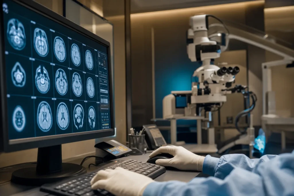
Meningioma
A meningioma is the most common tumour that forms inside the head, and the great majority are slow-growing and non-cancerous. Hearing the word "brain tumour" is frightening, but a meningioma is often very different from what that phrase brings to mind. This guide explains, in plain language, what a meningioma is, why it happens, how doctors find and treat it, and what life looks like afterwards, so you can have calmer, more informed conversations with your medical team.
What a meningioma is
A meningioma is a tumour (an abnormal growth of cells) that begins in the meninges — the thin protective layers of tissue that wrap around your brain and spinal cord, like a cover protecting the soft tissue underneath. More specifically, it grows from cells in the middle layer of this covering. Because it starts in the lining rather than in the brain tissue itself, a meningioma usually presses on the brain from the outside rather than growing through it.
This is an important distinction, and it is also reassuring for most people. A meningioma is the most common type of primary brain tumour (a tumour that starts in the head, rather than spreading there from somewhere else). According to the American Association of Neurological Surgeons, meningiomas make up roughly 38% of all primary central nervous system tumours. The large majority are benign, which means non-cancerous: they grow slowly, they do not normally invade the brain, and they very rarely spread to other parts of the body.
Many meningiomas grow so slowly, and cause so little trouble, that they are found completely by chance — for example, on a brain scan done for an unrelated headache or after a minor injury. A population study found meningiomas in around 1.8% of people scanned, often with no symptoms at all. So while "tumour in the brain" sounds alarming, for most people a meningioma is a slow, contained growth that can be watched or treated with good results.
Types and subtypes
Doctors describe meningiomas in two main ways: by where they are and by their grade (how the cells look and behave under a microscope).
By location. A meningioma can form almost anywhere the meninges exist. Cleveland Clinic describes common sites including convexity meningiomas (on the outer surface of the brain), sphenoid wing meningiomas (along a ridge of bone behind the eyes), olfactory groove meningiomas (between the brain and the nose), and intraventricular meningiomas (within the fluid-filled spaces of the brain). A small number form along the spinal cord. Location matters because it largely decides which symptoms, if any, appear, and how easy the tumour is to reach surgically.
By grade. The World Health Organization (WHO) sorts meningiomas into three grades:
- Grade 1 (typical): By far the most common. Slow-growing and non-cancerous.
- Grade 2 (atypical): Still usually non-cancerous, but grows faster and is more likely to come back after treatment. The AANS reports these make up roughly 18% of cases.
- Grade 3 (anaplastic or malignant): Rare — around 1.7% of meningiomas. These grow quickly and are cancerous.
The grade is decided by examining tumour tissue under a microscope, looking at how quickly the cells are dividing and whether they show certain features. Newer WHO guidance also takes genetic markers into account. The grade is one of the most useful pieces of information your team will have, because it guides both treatment and follow-up.
Causes and risk factors
For most people, the honest answer is that no one knows exactly why a meningioma formed. The NHS states plainly that the cause of most non-cancerous brain tumours is not known. What researchers have identified are risk factors — things that make a meningioma somewhat more likely. Having a risk factor does not mean you will get a meningioma, and many people with one have no risk factors at all.
The most consistently recognised factors include:
- Age. Risk rises with age. The AANS gives a median age at diagnosis of about 66 years, with risk increasing notably after 65.
- Sex and hormones. Meningiomas, especially grade 1, are more common in women. Researchers link this partly to the hormones oestrogen and progesterone. Cleveland Clinic notes that hormone replacement therapy, some birth control use, and a history of breast cancer have been associated with higher risk.
- Previous radiation to the head. People who had radiotherapy to the head in the past, sometimes for childhood conditions, have a higher long-term risk.
- Certain inherited conditions. Rare genetic syndromes such as neurofibromatosis type 2 and Li-Fraumeni syndrome increase risk. A family history of meningioma can also play a part.
At the cell level, Cleveland Clinic explains that most meningiomas involve a change (often the loss of part of a chromosome) in the genetic instructions of the cells. In the great majority of cases this change happens by chance during a person's lifetime and is not something you did or could have prevented.
Signs and symptoms, and when to see a doctor
Because a meningioma grows slowly and presses on whatever lies next to it, symptoms depend heavily on where the tumour is and how large it has become. Many small meningiomas cause no symptoms at all. When symptoms do appear, the NHS notes they usually build up gradually over months or even years, which is part of why they can be easy to dismiss at first.
Possible symptoms, drawn from Mayo Clinic and Cleveland Clinic, include:
- Headaches, sometimes worse in the morning
- Seizures (fits)
- Gradual changes in vision, such as blurred or double vision
- Hearing loss or loss of the sense of smell
- Weakness in an arm or a leg
- Memory problems, confusion, or changes in personality and mood
It is worth knowing that a tumour in the front part of the brain can occasionally show up first as low mood, anxiety, or personality change, which is why a thorough assessment matters when symptoms are unexplained.
When to see a doctor. See a doctor if you have headaches that are new, persistent, or getting worse, or that wake you from sleep; if you notice changes in vision, hearing, memory, or behaviour; or if you have any weakness or numbness. Seek urgent medical care if you have a first-ever seizure, sudden severe headache unlike any before, sudden confusion, or sudden loss of vision or movement. These symptoms have many possible causes — most are not a meningioma — but they always deserve prompt assessment.
Screening and early detection
There is no routine screening programme for meningioma. Because most are slow-growing, benign, and uncommon enough in the general population, there is no recommended scan or blood test for healthy people without symptoms. This is normal and reflects the nature of the condition rather than a gap in care.
In practice, a large share of meningiomas are found incidentally — that is, by accident — when a brain scan is done for some other reason, such as a head injury, dizziness, or migraines. When a small meningioma is discovered this way and is causing no problems, the usual response is careful monitoring rather than immediate treatment (more on this below).
People with a known higher risk — for example, those with neurofibromatosis type 2 or a strong family history of related tumours — may be offered regular monitoring by a specialist. If that applies to you, your genetics or neurology team will explain a personalised plan. For everyone else, the most useful form of "early detection" is simply not ignoring persistent or unusual symptoms and getting them checked.
How a meningioma is diagnosed
Diagnosis usually begins with a conversation about your symptoms and a neurological examination — simple checks of your vision, hearing, balance, strength, reflexes, and coordination that help the doctor work out which part of the brain might be affected.
The key test is brain imaging. As the NHS explains, an MRI (magnetic resonance imaging) scan is the main test; it uses magnets and radio waves, not radiation, to produce detailed pictures of the brain. A CT (computed tomography) scan, which uses X-rays, may be used as well, particularly to show calcium deposits or involvement of nearby bone. On these scans a meningioma often has a fairly characteristic appearance, and an experienced radiologist can frequently identify it with confidence. A contrast dye may be injected into a vein to make the tumour show up more clearly.
Imaging can strongly suggest a meningioma, but the only way to confirm the grade for certain is to examine the tumour tissue under a microscope. As the National Cancer Institute notes, this requires a sample of tissue, usually obtained during surgery to remove the tumour, which is then studied by a neuropathologist (a specialist in diagnosing nervous-system tumours). For small, symptom-free meningiomas that are being monitored rather than removed, this tissue diagnosis is often not needed unless the situation changes. Unlike some cancers, meningiomas are not "staged" with a number system; instead, the grade and the completeness of any surgery guide what happens next.
Treatment options
There is no single right treatment for a meningioma. The best choice depends on its size, location, grade, your symptoms, your age, and your overall health, and it is normally decided by a multidisciplinary team — typically a neurosurgeon, a radiation oncologist, a neurologist, a neuroradiologist, and a pathologist, working together. The main approaches are watchful waiting, surgery, and radiation.
Active monitoring (watch and wait). For small meningiomas that cause no symptoms, the safest option is often to do nothing immediately and instead watch the tumour with regular MRI scans, as Cancer Research UK and Mayo Clinic describe. Many such tumours grow so slowly that they never need treatment, and this spares people the risks of surgery.
Surgery. When a meningioma is causing symptoms or growing, surgery to remove it is the main treatment. Cleveland Clinic notes that a complete removal (called a gross total resection) can cure many meningiomas. Surgeons describe how completely a tumour was removed using the Simpson grading system, where lower numbers mean more complete removal and a lower chance of return. Sometimes, if a tumour is wrapped around important blood vessels or nerves, the surgeon will deliberately leave a small part behind to avoid harm, and treat the remainder another way.
Radiation therapy. Focused radiation can be used when surgery is not possible, when only part of the tumour could be removed, or for higher-grade tumours that are more likely to recur. A precise technique called stereotactic radiosurgery (for example, with a Gamma Knife) delivers a concentrated dose to the tumour in a way designed to spare surrounding tissue; despite the name, it does not involve cutting.
Supportive treatments. Medicines can manage symptoms — for example, anti-seizure medication, or steroids to reduce swelling around the tumour. Rehabilitation, such as physiotherapy or speech therapy, helps recovery. Chemotherapy has only a limited role and is generally reserved for rare tumours that keep coming back despite surgery and radiation.
Outlook: what to expect
For most people with a meningioma, the outlook is encouraging — but it genuinely depends on the grade, the location, and how completely the tumour can be treated. The figures below are population-level statistics from large groups of people. They describe averages and cannot predict what will happen for any one individual; your own outlook is something only your specialist can discuss with you in context.
Cancer Research UK reports, using data from England, that almost 70 out of 100 people (almost 70%) with a grade 1 or grade 2 meningioma survive for 10 years or more. For the rare grade 3 (malignant) meningiomas, the figure is around 40 out of 100 people (around 40%) surviving 10 years or more. The National Cancer Institute and Cleveland Clinic cite a five-year relative survival rate of 63.5% specifically for high-grade meningiomas. These numbers reflect older data and broad groups; survival statistics tend to improve over time as treatment advances, and they say nothing certain about a single person.
What most people can reasonably expect is this: a benign, fully removed grade 1 meningioma often does not return and may need only periodic check-ups. Higher-grade or incompletely removed tumours carry a greater chance of coming back and so call for closer follow-up and sometimes additional treatment. The key message is that grade and treatment options vary enormously between individuals, and a frank conversation with your own team will give you a far more meaningful picture than any general statistic.
Living with a meningioma and follow-up
Living well with a meningioma is very achievable for most people, but it usually involves ongoing follow-up rather than a single moment of "all clear." Because even benign meningiomas can sometimes return, regular monitoring is a standard and reassuring part of care, not a sign that something is wrong.
Follow-up scans. Whether you are being monitored without treatment or have already had surgery or radiation, you will normally have repeat MRI scans on a schedule set by your team — often more frequent at first, then spread further apart over the years if things are stable. The NHS notes that monitoring with regular scans may continue for several years.
Recovery and rehabilitation. After surgery, it is common to feel tired for a while, and some people experience temporary headaches, mood changes, or difficulty concentrating. Where the tumour or surgery affected a specific function — movement, speech, vision — rehabilitation therapies can help you regain ground. Recovery is gradual, and progress over weeks and months is the norm.
Day-to-day life. Practical points worth discussing with your doctor include whether and when you can drive (rules often apply after a seizure or brain surgery), returning to work, and managing any ongoing medication such as anti-seizure tablets. Many people return to full, active lives. Emotional support also matters: anxiety while waiting for scans (sometimes called "scanxiety") is common and normal, and talking to your team, a counsellor, or a patient support organisation can genuinely help.
Planning treatment abroad: what affects cost and how to prepare your records
If you are considering having a meningioma assessed or treated abroad, including in Turkiye, it helps to understand what shapes the overall cost and how to prepare. We do not publish fixed prices here, because every meningioma is different and a responsible estimate can only be made once specialists have reviewed your specific case. Instead, here is what tends to influence the cost and complexity of care.
- The treatment approach. Active monitoring, open surgery, and stereotactic radiosurgery are very different in scope, and each carries its own resource needs.
- Tumour location and complexity. A small, accessible meningioma is generally more straightforward than one near critical blood vessels, nerves, or deep structures, which may require longer, more specialised surgery.
- Grade and the need for added treatment. Higher-grade tumours may need radiation in addition to surgery, and closer follow-up.
- Diagnostic work-up. MRI, CT, and pathology all contribute, as may pre-operative assessments.
- Hospital stay and rehabilitation. The length of admission and any rehabilitation needs vary from person to person.
Preparing your records. To get an accurate, personalised opinion, gather your recent MRI and CT scans (ideally the actual image files on a disc or in digital form, not only the written report), any pathology results, a summary of your symptoms and their timeline, and a list of your medications and medical history. Having these ready allows specialists to give you meaningful guidance rather than a generic quote. The most reliable next step is to request a free consultation, share your records, and ask for a personalised estimate based on your own scans.
Why Turkiye, and how to choose a good centre
Turkiye has become a well-established destination for international patients seeking neurosurgery and related care, with a large number of internationally accredited hospitals and experienced specialist teams. For a condition like meningioma, where outcomes depend heavily on the skill of the surgical and radiation teams and on careful follow-up, choosing the right centre matters more than anything else. Rather than relying on marketing claims, focus on verifiable facts.
Things worth checking before you commit:
- Hospital accreditation. Joint Commission International (JCI) accreditation is a widely recognised international standard for patient safety and quality. You can confirm a hospital's status directly on the official Joint Commission International website rather than taking a third party's word for it.
- The specialist team. Ask who specifically will perform your surgery or radiation, their qualifications and experience with meningiomas, and whether a true multidisciplinary team (neurosurgery, radiation oncology, neuroradiology, pathology) reviews cases.
- The exact facility. Get the precise hospital name and address in writing before paying any deposit, and confirm that this is where your treatment will actually take place.
- Technology and follow-up. Check that the centre offers the imaging and, where relevant, the stereotactic radiosurgery equipment your case may need, and ask how follow-up scans and reports will be handled once you return home.
- Clear communication. A trustworthy centre will give you a written plan, an itemised personalised estimate, and access to interpreters, and will be transparent about what is and is not included.
A good concierge service should make these checks easier, not replace them. Use it to coordinate records, second opinions, and logistics, while you keep the final decision firmly in your own hands.
Second opinions, clinical trials, and looking after yourself
Because there is often more than one reasonable way to manage a meningioma — and because decisions about surgery versus monitoring versus radiation can be finely balanced — a second opinion is not only acceptable, it is sensible. Reputable specialists expect and welcome it. Sharing your scans and pathology with another qualified neurosurgical or neuro-oncology team can confirm a plan, surface alternatives, and give you confidence in whatever you decide.
For the small number of people with higher-grade, recurrent, or hard-to-treat meningiomas, clinical trials may be an option. These are carefully regulated studies testing newer approaches, such as targeted drugs or immunotherapy, that the National Cancer Institute notes are being investigated for meningioma. Trials are not right for everyone, and they are not a guarantee of benefit, but your team can tell you whether any are suitable and explain what taking part would involve.
Looking after yourself. There is no proven way to prevent a meningioma, since most arise by chance. What you can do is support your general health and your recovery: take any prescribed medication as directed, attend your follow-up scans, avoid known triggers for seizures if you have had one (such as lack of sleep), stay physically active as your team advises, and reach out for emotional support when you need it. Above all, stay connected with a qualified specialist who knows your case — that ongoing relationship is the single most valuable thing for managing a meningioma well over time.
Frequently asked questions
Is a meningioma cancer?
Does every meningioma need to be removed?
What are the first signs of a meningioma?
What causes a meningioma?
How is a meningioma diagnosed?
Can a meningioma come back after treatment?
What is the outlook for someone with a meningioma?
Is there screening for meningioma?
What does treatment cost in Turkiye?
How do I choose a hospital in Turkiye for meningioma treatment?
Should I get a second opinion?
This article is for general information only and is not medical advice. Always consult a qualified doctor about your individual case.
Considering this procedure?
Send us your photos and questions. A BergemHealth coordinator and a department-head specialist will review your case and reply with honest, personalised guidance — no obligation.
Free consultation