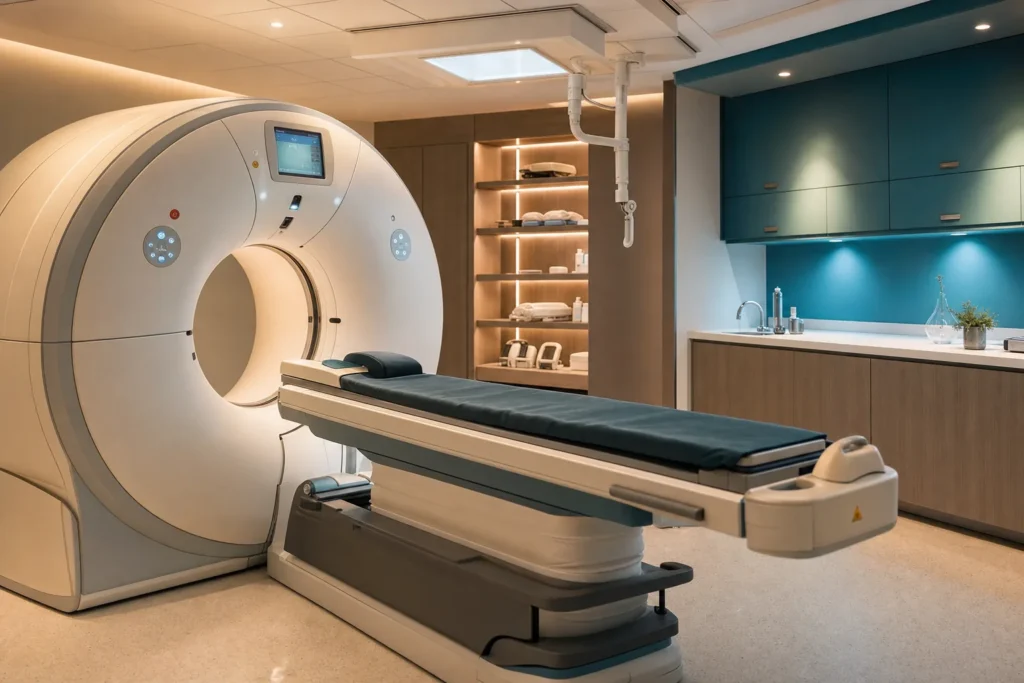
PET/CT scan
A PET/CT scan is one of the most informative pictures modern medicine can take of the inside of your body. Unlike an ordinary X-ray or scan that shows only shape and structure, a PET/CT shows how busy your cells actually are, which can reveal disease before it changes a tissue's outline. This guide explains in plain words what the scan is, who it helps, how the day unfolds, what is safe and what is not, how long results take, and what it tends to cost if you arrange it in Turkiye.
- Anaesthesia
- None needed; a light sedative is occasionally offered for severe claustrophobia.
- Duration
- About 2 to 3 hours total: roughly 1 hour resting after the tracer injection, then 15 to 45 minutes of scanning.
- Recovery
- Immediate; you can resume normal activities the same day. Drink plenty of water to clear the tracer.
- Hospital stay
- None; it is an outpatient (day) test with no overnight stay.
Indicative hospital range, confirmed after your file is reviewed. You pay the hospital’s price — we add no markup.
What a PET/CT scan is
A PET/CT scan is two scans combined into one machine and one appointment. The letters stand for two different imaging methods working together:
- PET means positron emission tomography. It shows how active your cells are by tracking a small amount of a radioactive substance you are given. Busy, energy-hungry cells light up; quiet ones stay dim.
- CT means computed tomography. It is a fast series of X-rays that builds a detailed map of your anatomy, the actual shape and position of your organs, bones and soft tissues.
On their own, each test has a weakness. PET shows activity but is fuzzy about exact location. CT shows precise location but cannot tell whether a lump is alive and active or just old scar tissue. Put together, a PET/CT overlays the two, so a doctor can see a bright "hot spot" of activity and pinpoint exactly which organ or lymph node it sits in.
The radioactive substance is called a radiotracer (or tracer). The most common one is FDG, short for fluorodeoxyglucose, which is essentially a form of sugar with a tiny radioactive tag attached. Because cells use sugar for fuel, and many cancer cells are unusually hungry for it, they soak up more FDG than normal tissue and show up brightly. According to RadiologyInfo (from the Radiological Society of North America), PET is a type of nuclear medicine imaging that lets doctors look at function at the cellular level, sometimes spotting a problem earlier than tests that only show structure.
Who is a good candidate (and who should avoid it)
A PET/CT is not a routine check-up scan for everyone. It is ordered for specific questions, most often around cancer, but also for heart and brain conditions. Common reasons include:
- Cancer: finding out whether a lump is likely cancerous, checking whether a known cancer has spread (called staging), seeing whether treatment is working, and telling active cancer apart from leftover scar tissue after treatment.
- Heart disease: assessing blood flow to the heart muscle and whether damaged areas might recover.
- Brain conditions: helping investigate some forms of dementia, and helping locate the source of seizures when planning epilepsy surgery.
Your own doctor decides whether the scan will genuinely answer the question being asked. Some people should usually avoid a PET/CT or need special planning:
- Pregnancy: the NHS and Cancer Research UK advise that scans are generally avoided in pregnancy and only done in an emergency, because radiation can harm an unborn baby. Always tell the team if you are or might be pregnant.
- Breastfeeding: the scan can still be done, but you will usually be asked to pause breastfeeding for a few hours afterwards (Memorial Sloan Kettering advises stopping for about 4 hours and discarding or storing pumped milk).
- Poorly controlled diabetes: high blood sugar competes with the FDG tracer and can blur the result. If your blood glucose is too high on the day (typically 200 mg/dL or above), the scan may be rescheduled.
- Severe claustrophobia or difficulty lying still: manageable, but worth flagging in advance.
- Very large body size: there are weight and width limits for the scanner, so the clinic may need to check the equipment can accommodate you.
Types and techniques
Most PET/CT scans use the same general method, but the tracer can be chosen to match the question. This is where the test becomes tailored:
- FDG (fluorodeoxyglucose): the workhorse tracer, a radioactive sugar used for most cancer scans and for many heart and brain studies. It is made in a machine called a cyclotron and has a half-life of about 110 minutes, meaning it loses half its radioactivity in under two hours, so timing matters.
- Gallium-68 PSMA: targets a protein found on prostate cancer cells, used to stage prostate cancer and look for spread.
- Gallium-68 DOTATATE (or DOTATOC): attaches to receptors on certain rare tumours called neuroendocrine tumours, and is a leading way to image them.
A practical difference: gallium-68 tracers can be made on site with a small generator, so a clinic does not need its own cyclotron nearby, whereas FDG must be delivered fresh because it decays quickly.
You may also hear about whole-body versus targeted scans. A whole-body PET/CT typically images from the base of the skull to the upper thighs, while a dedicated brain or heart study focuses on one region. The CT part can be done as a low-dose scan purely to map location, or as a fuller diagnostic CT, sometimes with a separate contrast dye, which affects both the information gained and the radiation dose.
How it is done: anaesthesia, steps and timing
A PET/CT needs no anaesthesia. You stay awake throughout. The only injection is the tracer itself, given through a small drip in your arm. People with severe claustrophobia are occasionally offered a mild sedative, but most need nothing because the scanner is an open ring rather than a long enclosed tunnel.
Here is how a typical FDG appointment unfolds:
- Check-in and blood sugar test. A finger-prick or small blood sample checks your glucose level, because high sugar can spoil the images.
- Tracer injection. A nurse or technologist places a thin cannula in a vein and injects the FDG. It is painless apart from the small needle.
- The quiet wait (uptake period). You rest in a calm room for about 60 minutes while your body takes up the tracer. You are asked to stay relaxed and avoid talking, chewing or moving much, because active muscles also absorb sugar and can create misleading bright areas. You may be given a drink of oral contrast during this time.
- The scan. You lie on a bed that slides slowly through the doughnut-shaped scanner. The CT part is very quick (often under two minutes) and the PET part takes longer. Overall scanning usually lasts 15 to 45 minutes depending on the type of study; it is painless and you simply need to lie still.
Counting the rest period, scan and paperwork, plan for roughly two to three hours at the clinic. The actual machine time is a small part of that.
Recovery, step by step
Recovery from a PET/CT is straightforward because nothing is cut, stitched or removed. There is no wound and no stitches.
- Straight after: the cannula is removed and you can get dressed and go. Most people return to their usual day, including eating, drinking and driving, unless they were given a sedative.
- The first few hours: drink plenty of water. The tracer leaves your body naturally, mainly through urine, and fluids help flush it out faster.
- Radiation precautions for about a day: for a short period you are very mildly radioactive. As a sensible precaution, the NHS and major cancer centres advise avoiding prolonged close contact with young children and anyone who is pregnant for the rest of the day. Memorial Sloan Kettering suggests avoiding skin-to-skin contact and sharing a bed with pregnant people or under-18s for the first hours after the scan.
- Injection site: keep an eye on where the cannula went in for a day or two. A small bruise is normal; redness, swelling, increasing pain or discharge are not, and should be reported.
- Breastfeeding: if this applies, follow the team's advice on pausing feeds, commonly for about four hours.
There is no formal "recovery period" in the surgical sense. By the next day, any precautions have passed.
Risks and possible complications
A PET/CT is considered a low-risk test, and reputable sources stress that for the people who need it, the benefit of an accurate diagnosis far outweighs the small risks. The main considerations are:
- Radiation exposure. Both the tracer and the CT involve a small dose of radiation. The dose adds slightly to your lifetime radiation total and, in theory, to a very small long-term risk; in practice it is treated as low and acceptable for a diagnostic test. A typical whole-body FDG PET/CT delivers in the region of about 10 to 25 millisieverts depending on the CT settings used, compared with average natural background radiation of roughly 2.4 to 3 millisieverts a year that everyone receives from the environment. Doctors have used nuclear medicine scans for more than six decades.
- Injection site reactions. As with any blood test, there is a small chance of bruising, soreness, bleeding or, rarely, infection where the needle goes in.
- Allergic reaction. A true allergic reaction to the PET tracer is very rare and usually mild, because the amount used is tiny. If a separate CT contrast dye is used, that carries its own small, separate reaction risk, which the team will ask about beforehand.
- Discomfort or anxiety. Lying still, or being in the scanner, can be uncomfortable or trigger claustrophobia in some people.
- Inaccurate results. High blood sugar, recent vigorous exercise, infection or inflammation can all create bright spots that mimic or hide disease, which is why preparation rules matter.
There is no general risk to people who care for you afterwards, beyond the simple precautions about close contact for the rest of the day.
Results and how long they last
A PET/CT is a diagnostic test, not a treatment, so it does not produce a result that "lasts" in the way surgery does. Instead, it produces a snapshot of how your body looked on that day.
The images are reviewed by a specialist doctor (a nuclear medicine physician or radiologist) who writes a report for the doctor who referred you. The NHS and Cancer Research UK note it usually takes about one to two weeks to get your results, because the images are studied carefully and often discussed alongside your other tests and scans.
How long the information stays useful depends on why the scan was done. For staging a cancer, the snapshot guides decisions made over the following weeks. For checking whether treatment is working, doctors often repeat the scan later to compare. Because disease can change, a PET/CT reflects a moment in time rather than a permanent answer, and your team will decide if and when another scan is worthwhile.
Costs: indicative ranges and what changes the price
Prices for a PET/CT in Turkiye are generally lower than in many Western European countries, which is one reason people travel for the test. As an indicative guide, a PET/CT scan in Turkiye commonly falls in the region of about €500 to €1,500, with whole-body FDG studies clustering in the middle of that range and specialised tracers at the higher end.
These figures are an indication only. The price varies by case, by clinic and by the doctors involved, and is not a quote. Several things move the number up or down:
- The tracer used. Standard FDG is the most affordable. Specialised tracers such as gallium-68 PSMA (for prostate cancer) or DOTATATE (for neuroendocrine tumours) usually cost more.
- Scope of the scan. A whole-body study costs more than a single-region scan, and a full diagnostic CT with contrast dye adds to the price compared with a low-dose CT used only for location.
- The clinic and equipment. Internationally accredited hospitals with newer scanners may price differently from smaller centres.
- What is included. Some prices cover only the scan, while packages may include the specialist's report, a consultation, or interpreter and transfer services for international patients.
When comparing offers, always ask exactly what is and is not included, so you are comparing like with like.
Why people travel to Turkiye, and how to choose a safe clinic
Turkiye has become a well-known destination for medical imaging and treatment because it combines modern hospitals, experienced specialists and prices that are often lower than in Western Europe. Many of its hospitals hold international accreditation and use current-generation scanners.
A scan is only as good as the people reading it, so choosing carefully matters. Practical checks before you book:
- Accreditation. Look for hospitals accredited by Joint Commission International (JCI), a widely respected global standard for quality and patient safety. Turkiye has many JCI-accredited hospitals. You can confirm a hospital's current status on the official Joint Commission International website rather than relying only on the clinic's own claims, and check that the exact facility name and location match.
- Who reads the scan. The images should be reported by a qualified nuclear medicine physician or radiologist. Ask whether you will receive a written report, in which language, and whether you can get the raw images on a disc or digitally to share with your own doctor at home.
- The equipment and tracer. Ask which tracer will be used and whether it suits your specific question, and confirm the clinic can supply it (especially for non-FDG tracers).
- Clear, written pricing. A trustworthy clinic gives an itemised quote and does not promise specific findings or outcomes in advance.
- Continuity of care. Consider how the result will reach the doctor managing your treatment, and how follow-up questions will be handled once you are home.
Be cautious of any clinic that guarantees a particular result or pressures you to decide quickly.
How to prepare and what to ask at your consultation
Good preparation directly affects how reliable your scan is. For a standard FDG PET/CT, typical instructions (based on NHS, Cancer Research UK and Memorial Sloan Kettering guidance) include:
- Fasting: do not eat for about 4 to 6 hours beforehand. You may usually drink plain water, but avoid sugary or milky drinks, gum, mints and sweets, which add sugar that competes with the tracer.
- Avoid hard exercise for 24 hours before, since active muscles soak up sugar and can blur results.
- Manage diabetes carefully: ask your doctor how to handle your medication and blood sugar on the day, as the scan may be postponed if your glucose is too high.
- Stay warm: some clinics ask you to keep warm, because cold can activate certain tissues that then show up on the scan.
- Remove metal: jewellery and metal items are removed before scanning.
- Tell the team if you are or might be pregnant, are breastfeeding, are claustrophobic, have allergies, or wear an insulin pump or continuous glucose monitor.
Helpful questions to ask at your consultation:
- Which tracer will be used, and why is it right for my situation?
- Will this be a whole-body or targeted scan, and will contrast dye be used?
- How should I prepare, and what should I do about my regular medicines?
- How long will the whole appointment take?
- When and how will I get my results, and in what language is the report?
- Can I take the images home to share with my own doctor?
Aftercare and travelling for the test (including flying)
Because a PET/CT is an outpatient test with no surgery, aftercare is simple. Drink plenty of fluids for the rest of the day to help clear the tracer, keep your distance from young children and anyone pregnant for that day, and watch the injection site for a couple of days for any unusual redness or swelling.
Is it safe to fly afterwards? Medically, yes. By the time you leave the clinic the tracer is already fading, and you can usually travel the same day or the next day. There is no surgical wound to worry about, so the flying precautions that apply after operations do not apply here. There is one practical wrinkle for international patients: for a short time after the scan you may carry enough residual radioactivity to trigger sensitive radiation detectors at airports. This is harmless, but if you are flying soon after the scan, ask the clinic for a letter or card stating that you have had a nuclear medicine test, so you can show it to security if needed.
If you are travelling to Turkiye specifically for the scan, plan a little buffer time in case your appointment is rescheduled (for example, if your blood sugar is high on the day) and to allow the specialist to prepare your report. Arrange in advance how the images and report will reach the doctor managing your care back home, ideally as both a written report and a digital copy of the images, so the journey translates into useful answers rather than just a scan.
Frequently asked questions
Does a PET/CT scan hurt?
Do I need to be put to sleep or sedated?
How long does the whole appointment take?
Why do I have to fast and avoid sugar before the scan?
Is the radiation dangerous?
Am I radioactive afterwards, and is it a risk to my family?
Can I drive and go back to normal life the same day?
Can I have a PET/CT if I am pregnant or breastfeeding?
When will I get my results?
How much does a PET/CT scan cost in Turkiye?
Is it safe to fly soon after a PET/CT?
Is FDG the only tracer used?
This article is for general information only and is not medical advice. Always consult a qualified doctor about your individual case.
Considering this procedure?
Send us your photos and questions. A BergemHealth coordinator and a department-head specialist will review your case and reply with honest, personalised guidance — no obligation.
Free consultation