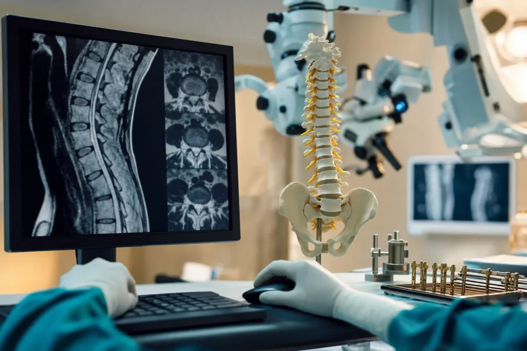
Spinal cord tumours
A spinal cord tumour is an abnormal growth of cells in or around the spinal cord. The words sound frightening, but most spinal tumours are not cancer, many are highly treatable, and modern neurosurgery and imaging have made careful, organ-sparing treatment possible. This guide explains, in plain language, what spinal cord tumours are, the warning signs worth knowing, how doctors find and treat them, and how to prepare if you are considering care abroad.
What a spinal cord tumour is
Your spinal cord is a soft, ribbon-like bundle of nerves that runs from the base of your brain down through the bones of your spine (the vertebrae). It carries the signals that let you move, feel and control your bladder and bowel. A spinal cord tumour is an abnormal growth of cells inside or around this cord and the column of bone that protects it.
Doctors describe these growths in two important ways. First, by whether they are benign or malignant. Benign means non-cancerous: the growth does not spread to other parts of the body, though it can still cause problems by pressing on the cord. Malignant means cancerous: the cells can grow into nearby tissue and, in some cases, spread elsewhere.
Second, doctors describe a tumour by where it sits. According to the Cleveland Clinic, growths outside the spinal cord's covering, often in the bones of the spine, make up roughly 55% of cases; growths inside the covering but outside the cord itself make up about 40%; and growths inside the cord tissue are the least common, at around 5%. Most spinal tumours overall are not growths that started in the spine at all, but cancer that has travelled there from elsewhere in the body (covered below).
It helps to keep perspective. Primary spinal cord tumours, ones that begin in the spine or cord, are uncommon. Cancer Research UK notes they account for only about 3 in every 100 brain and spinal tumours. Knowing what you are dealing with is the first step toward calm, clear decisions.
Types and subtypes
Spinal cord tumours are usually grouped by their location, because location strongly affects symptoms and treatment. The three main groups are:
- Extradural (or epidural) tumours sit outside the dura, the tough membrane that wraps the cord. They most often involve the bones of the spine. This is the group most commonly linked to cancer that has spread from elsewhere.
- Intradural-extramedullary tumours sit inside the dura but outside the cord itself. Common examples include meningiomas (growths of the cord's covering), schwannomas and neurofibromas (growths arising from the nerve sheaths). These are very often benign.
- Intramedullary tumours grow within the cord tissue itself. The two most common are ependymomas (from the cells lining the cord's fluid channels) and astrocytomas (from supporting cells called glia). A less common type, haemangioblastoma, arises from blood-vessel cells.
Tumours are also given a grade, a measure of how the cells look under the microscope and how quickly they tend to grow. Low-grade means slow-growing; high-grade means faster-growing. As Cancer Research UK explains, grade is one of several factors that shape treatment and outlook.
A separate, very important category is metastatic (secondary) spinal tumours, cancer cells that have spread to the spine from a cancer that began somewhere else, such as the breast, lung, prostate, kidney or thyroid. These are the most common spinal tumours in adults and are managed somewhat differently from primary tumours.
Causes and risk factors
For most people, the honest answer is that the exact cause of a primary spinal cord tumour is not known. Tumours form when cells acquire errors in the genes that control how they grow and divide, but why this happens in a particular person is usually unclear. Importantly, nothing you did caused it.
A small number of people have an inherited condition that raises the risk. Mayo Clinic and the Cleveland Clinic list the main ones:
- Neurofibromatosis type 1 (NF1) and type 2 (NF2) — inherited conditions that predispose to growths on nerves and nerve coverings.
- Von Hippel-Lindau disease (VHL) — an inherited condition linked to blood-vessel tumours (haemangioblastomas) in the spinal cord, brain and retina, among other growths.
People with a weakened immune system have a higher chance of a rare spinal lymphoma. For metastatic spinal tumours, the risk factor is simply having a cancer elsewhere in the body that can spread to bone, most commonly breast, lung, prostate, kidney, thyroid, gastrointestinal cancers and multiple myeloma.
It is worth saying clearly: the great majority of people with a spinal tumour have no identifiable risk factor at all. Having one of these conditions does not mean a tumour is certain, and not having them does not make you immune. If a hereditary syndrome runs in your family, your doctor can advise whether monitoring makes sense.
Signs and symptoms, and when to see a doctor
Symptoms depend on where the tumour is and what it is pressing on. Because they often come on gradually, they can be mistaken for ordinary back trouble. The most common first symptom, described by both the Cleveland Clinic and specialist reviews, is back or neck pain. Certain features make tumour-related pain more concerning:
- It is not linked to an injury or to activity.
- It is deep and aching, and tends to get worse over time.
- It is often worse at night or at rest and may disturb sleep.
- It does not improve with usual painkillers or rest.
Other possible signs, especially as a tumour presses on nerves, include:
- Pain that shoots from the spine into the arms, legs, hips or feet.
- Numbness, tingling or reduced sensation to touch, heat or cold.
- Muscle weakness, clumsiness, or difficulty walking and keeping balance.
- Reduced control of the bladder or bowel.
Please see a doctor promptly if you have back pain with any of these features, or new neurological symptoms. Most back pain is not a tumour, but it is always reasonable to have unexplained, persistent or night-time pain checked.
Seek urgent or emergency care if you develop rapidly worsening leg weakness, numbness spreading in the legs or around the groin and buttocks, or new loss of bladder or bowel control. In someone with a known cancer, this combination may signal spinal cord compression, which the NHS and NICE treat as a true emergency. As guidance summarised by NICE explains, prompt MRI (ideally within 24 hours) and treatment give the best chance of preserving nerve function.
Screening and early detection
There is no routine screening test for spinal cord tumours in the general population. Because primary spinal tumours are rare and there is no simple, reliable test that finds them early in healthy people, screening everyone would not be useful. This is normal for many uncommon conditions and is not a gap in your care.
The exceptions are people with an inherited condition known to raise the risk, such as von Hippel-Lindau disease or neurofibromatosis. For them, specialists may recommend periodic check-ups and scans of the nervous system to catch growths early. If you have such a diagnosis, your medical team will explain a monitoring plan tailored to you.
For everyone else, "early detection" really means not ignoring persistent warning signs. Pain that does not behave like ordinary muscle strain, especially night pain, or any new weakness, numbness or bladder and bowel change, deserves medical assessment. In people with a known cancer, doctors stay alert for symptoms of spread to the spine, which is one reason follow-up after a cancer diagnosis is important.
How a spinal cord tumour is diagnosed
Diagnosis usually begins with a conversation about your symptoms and a neurological examination, in which the doctor checks your strength, reflexes, sensation, balance and bladder or bowel function. This helps locate where in the spine a problem might be.
Imaging is the key step. Magnetic resonance imaging (MRI) is described by the NINDS and specialist sources as the gold-standard test. It uses a strong magnet and radio waves (no radiation) to produce detailed pictures of the cord, nerves and surrounding tissue. A contrast dye is often given through a vein to make a tumour stand out and show its size, position and any associated swelling or fluid-filled cysts. A CT scan may be added to look at the bones in detail.
To know exactly what type of tumour it is, doctors usually need a biopsy, a small sample of tissue examined under a microscope. This is sometimes taken with a needle and sometimes during surgery to remove the tumour. The biopsy reveals whether the growth is benign or malignant, its specific type and its grade, all of which guide treatment.
If a tumour looks like it may have spread from elsewhere, further tests, such as scans of the chest and abdomen, blood tests, or a bone scan, help find the original cancer. For intramedullary tumours, MRI features can hint at the type: ependymomas often sit centrally and have a clear edge, while astrocytomas tend to be more off-centre with less defined borders.
Treatment options
Treatment is always individualised, and it is usually planned by a multidisciplinary team, a group that may include a neurosurgeon, a neuro-oncologist (cancer specialist), a radiation oncologist, a radiologist, a pathologist, and rehabilitation specialists such as physiotherapists. They weigh the tumour type, grade, location and your overall health to recommend an approach. The main options are:
- Watchful waiting (monitoring). Small, slow-growing or symptom-free benign tumours, especially some that sit inside the dura but outside the cord, may simply be watched with regular MRI scans rather than treated straight away. As the AANS notes, surgery on the cord itself carries some risk, so observation is sometimes the wisest first step.
- Surgery. For many tumours, an operation to remove as much of the growth as safely possible is the central treatment. Microsurgical techniques and intraoperative monitoring (watching nerve signals during the operation) help the surgeon protect the cord. For some clearly bordered tumours, such as many ependymomas, complete removal is often achievable; for tumours that blend into the cord, such as some astrocytomas, surgeons may remove part of the tumour to relieve pressure while avoiding nerve damage.
- Radiation therapy. High-energy beams can shrink or control tumour that cannot be fully removed, or treat higher-grade tumours after surgery. Stereotactic radiosurgery delivers a precise, focused dose, and is often used for metastatic tumours.
- Chemotherapy. Medicines that kill or slow cancer cells are used in selected cases. Cancer Research UK notes chemotherapy is used less often for primary spinal cord tumours because it tends to be less effective there than for some brain tumours.
- Supportive treatment. Steroids are commonly given to reduce swelling around the cord and ease symptoms, and other medicines manage pain. Procedures that stabilise weakened spinal bones can relieve pain in metastatic disease.
Outlook: what to expect
It is natural to want a clear answer about the future, but the honest picture is that outlook varies a great deal from person to person. As the NINDS and Cleveland Clinic both emphasise, prognosis depends on the type and grade of the tumour, its location, your age and general health, and how much can be safely treated. No article, and no statistic, can predict what will happen for any one individual.
A few general patterns are well established. Many benign tumours, particularly meningiomas and schwannomas that can be fully removed, are associated with good outcomes once treated. For intramedullary tumours, specialist reviews note that the strongest predictor of how well someone does afterward is their neurological condition before surgery, which is one of the reasons doctors encourage early assessment rather than waiting. Clearly bordered ependymomas can often be removed completely; infiltrating astrocytomas are more challenging.
Where survival figures exist, they come from large groups of patients and describe averages, not predictions. They cannot tell you, personally, what to expect. The most reliable source of information about your own situation is the specialist who has reviewed your scans and pathology. It is completely reasonable to ask them, in plain terms, what the realistic goals of your treatment are.
Living with a spinal cord tumour and follow-up
Recovery and daily life after treatment depend on how the tumour and its treatment affected your nerves. Many people regain function, and rehabilitation is a central part of care. Physiotherapy helps rebuild strength, balance and walking; occupational therapy helps with everyday tasks; and specialists can advise on managing nerve pain, bladder and bowel changes, and fatigue. Starting rehabilitation early, where appropriate, supports the best functional recovery.
Follow-up usually involves regular clinic visits and repeat MRI scans to check that a treated tumour has not returned or grown, and to watch tumours that are being monitored. The schedule depends on the tumour type and grade. After cancer treatment generally, follow-up also helps detect any new spread to the spine early.
Living well also means caring for the parts of life beyond the scans. Pacing activity, gentle exercise as advised, good sleep, and support for low mood or anxiety all matter. Many people find peer-support groups and patient organisations valuable. Tell your team promptly about any new or worsening pain, weakness, numbness, or bladder and bowel changes, rather than waiting for the next appointment.
Planning treatment abroad: what affects cost and how to prepare your records
If you are considering treatment in another country, it helps to understand what shapes the overall cost, so you can plan and compare fairly. We do not list prices here because every case is genuinely different; instead, ask for a personalised estimate after a specialist has reviewed your records. The main factors include:
- The tumour and the plan. Type, grade and location determine whether treatment is surgery, radiation, a combination, or monitoring, and how complex any operation is (for example, intramedullary surgery with intraoperative nerve monitoring).
- Diagnostics. Whether new MRI, CT, biopsy or laboratory work is needed, or whether your existing scans can be reviewed.
- Hospital stay and intensive care needs, and any rehabilitation or physiotherapy afterward.
- Implants and equipment if spinal stabilisation is required.
- Follow-up scans, medicines and clinic reviews.
- Travel and accommodation for you and a companion, and translation or coordination support.
To get an accurate estimate and avoid repeating tests, gather your records before you reach out: recent MRI and CT images (the actual files, not just the report), radiology and pathology/biopsy reports, a summary letter from your current doctor, a list of medicines and allergies, and relevant blood test results. A specialist can then review them remotely and tell you what is realistic. A free consultation is the simplest way to get a tailored estimate and a clear plan.
Why Turkiye, and how to choose a good centre
Turkiye (Turkey) has become a well-established destination for neurosurgical and cancer care, with many internationally accredited hospitals and experienced teams, often at a lower cost than in Western Europe or North America. The point of seeking care abroad is not to find a "famous" name but to find a centre that is genuinely well suited to your diagnosis. Here is what to verify:
- International accreditation. Look for hospitals accredited by Joint Commission International (JCI), a global standard for patient safety and quality. Turkiye is among the countries with the most JCI-accredited facilities; you can confirm a hospital's status on the official Joint Commission International register.
- A dedicated neurosurgical and multidisciplinary team. Spinal cord tumour care should involve neurosurgeons experienced specifically in spinal cord (not just spine bone) surgery, working alongside neuro-oncology, radiation oncology, radiology, pathology and rehabilitation. Ask how many such cases the team handles.
- Modern equipment. High-quality MRI, the option of stereotactic radiosurgery where appropriate, and intraoperative neuromonitoring during surgery are important markers of a capable centre.
- Clear communication. A good centre will offer a written treatment plan, explain risks and goals honestly, provide your records and pathology results, and arrange interpreters and a coordinator for international patients.
- Realistic claims. Be cautious of any provider promising a guaranteed cure or using "best" or "number one" language. Reputable teams discuss goals and probabilities, not certainties.
It is always reasonable, and often wise, to seek a second opinion before committing to a plan. A trustworthy centre will welcome that.
Clinical trials and getting a second opinion
There is no proven way to prevent most spinal cord tumours, so the most useful self-care is staying informed and proactive about your own care. Two avenues are worth knowing about.
First, clinical trials. Research is active in this field, including newer approaches such as targeted therapies and immunotherapies for certain tumour types, as noted by the NINDS. Trials are not a last resort; for some people they offer access to promising treatments under careful supervision. Ask your specialist whether any trial is appropriate for your tumour type and grade, and what it would involve.
Second, a second opinion. Because spinal cord tumours are uncommon and treatment decisions are nuanced, having another qualified specialist review your scans and pathology can confirm the diagnosis, clarify the options, and give you confidence in the plan. This is a normal, accepted part of good care, not a sign of distrust.
Finally, look after the basics that support any recovery: keep up with follow-up appointments, take medicines as prescribed, stay as active as your team advises, eat well, sleep, and seek support for the emotional side of a diagnosis. If you have an inherited condition that raises your risk, follow the monitoring plan your specialists recommend. And whenever new neurological symptoms appear, have them checked promptly, early assessment consistently gives the best chance of protecting function.
Frequently asked questions
Is a spinal cord tumour the same as cancer?
Are spinal cord tumours common?
What is usually the first symptom?
When is back pain an emergency?
How are spinal cord tumours diagnosed?
Can a spinal cord tumour be removed completely?
Is surgery always needed?
What is the outlook after treatment?
Will I be able to walk and function normally afterward?
What records should I gather before seeking treatment abroad?
How do I choose a good hospital in Turkiye?
Are there inherited conditions that raise the risk?
This article is for general information only and is not medical advice. Always consult a qualified doctor about your individual case.
Considering this procedure?
Send us your photos and questions. A BergemHealth coordinator and a department-head specialist will review your case and reply with honest, personalised guidance — no obligation.
Free consultation