Bronchiectasis
Bronchiectasis is a long-term lung condition in which some of your airways become permanently widened and find it harder to clear mucus. It cannot be cured, but for most people it can be managed well, and many live full and active lives. This guide explains in plain words what bronchiectasis is, why it happens, how it is diagnosed and treated, and how to prepare if you are considering care abroad.
What bronchiectasis is
Bronchiectasis (say it "bron-kee-EK-ta-sis") is a long-term condition in which one or more of the airways in your lungs become permanently widened. Your airways are the branching tubes, called bronchi, that carry air in and out of your lungs. In bronchiectasis the walls of some of these tubes are damaged by inflammation (the body's swelling and irritation response), so they stretch wider than they should and become floppy.
Healthy airways are lined with tiny hair-like brushes called cilia that constantly sweep mucus (the slippery fluid that traps dust and germs) up and out of the lungs. When the airways are widened and damaged, this cleaning system stops working properly. Mucus then pools in the floppy areas instead of being cleared, and germs can grow in it. This leads to a long-lasting (chronic) cough that brings up phlegm, and to repeated chest infections.
Doctors often describe bronchiectasis as a "vicious cycle." Mucus that is not cleared invites infection; infection causes more inflammation; inflammation causes more airway damage; and the more damaged the airway, the harder it is to clear mucus. Treatment is aimed at breaking this cycle. It is important to understand that the widening of the airways is permanent and cannot be reversed, but with the right care the cycle can be slowed or quietened, and symptoms kept under control.
Bronchiectasis is not the same as bronchitis, asthma or chronic obstructive pulmonary disease (COPD), although it can occur alongside them. It is more common in older adults and is reported slightly more often in women than in men.
Types and subtypes
There is no single "type" of bronchiectasis. Doctors usually describe it in two practical ways: by what the damaged airways look like on a scan, and by how much of the lung is affected.
By shape of the airway. Specialists may use these words when reading a CT scan:
- Cylindrical (tubular): the airways are widened evenly, like a smooth tube. This is the most common pattern and usually the mildest.
- Varicose: the airways are wider in some places and narrower in others, a bit like beads on a string.
- Cystic (saccular): the airways form balloon-like pouches or honeycomb-shaped pockets. This is usually the most advanced pattern.
These descriptions help your team understand the extent of damage, but they do not by themselves decide your treatment.
By how much lung is involved. Bronchiectasis may be focal (limited to one part of one lung, often after a single past problem such as a blockage or one severe infection) or diffuse (scattered across several areas of both lungs, more often linked to a body-wide cause). Focal disease in one small area is occasionally suitable for surgery; diffuse disease is managed with medicines and airway-clearance techniques.
You may also see the term traction bronchiectasis, where scarring in the surrounding lung tissue pulls the airways open. A very common everyday distinction is between bronchiectasis caused by cystic fibrosis (an inherited condition) and non-cystic-fibrosis bronchiectasis, which covers everyone else and is what this guide mainly describes.
Causes and risk factors
Bronchiectasis develops when something damages the airways. Sometimes a single event is responsible; sometimes an ongoing condition keeps the airways inflamed. In a large share of cases no clear cause is ever found even after thorough testing. The American Lung Association notes that the cause is unknown in roughly 40% of people.
Recognised causes and contributing factors include:
- A previous severe lung infection, such as pneumonia, whooping cough (pertussis), measles, tuberculosis (TB) or, more recently, COVID-19. The infection may have happened years earlier, even in childhood.
- Other lung conditions, including asthma and COPD.
- Cystic fibrosis, an inherited condition that causes thick, sticky mucus throughout the body.
- Problems with the immune system, where the body cannot fight infections normally, so chest infections recur.
- Primary ciliary dyskinesia, an inherited problem in which the cilia (the airway's cleaning brushes) do not work properly.
- Allergic bronchopulmonary aspergillosis, an allergic reaction in the lungs to a common mould.
- Inflammatory and autoimmune conditions, such as rheumatoid arthritis, Sjogren syndrome, ulcerative colitis or Crohn's disease.
- A blockage in an airway, for example an inhaled object, an enlarged lymph node or a growth.
- Infection with non-tuberculous mycobacteria, a group of germs related to TB.
Risk rises with age, and finding an underlying cause matters because treating it (for example, an immune problem or an allergy) can help protect the lungs from further harm.
Signs and symptoms, and when to see a doctor
The most common symptom of bronchiectasis is a cough that lasts for months or years and brings up phlegm (sputum). The amount of phlegm varies a lot between people: some bring up only a little, while others cough up a cupful or more over a day. The phlegm may be clear, white, yellow or green, and it can sometimes smell unpleasant.
Other common symptoms include:
- Repeated chest infections that take longer than usual to clear.
- Feeling very tired or low on energy.
- Shortness of breath, particularly during activity.
- Wheezing (a whistling sound when you breathe).
- Chest pain, which can be sharper during an infection.
- Coughing up small amounts of blood, or phlegm streaked with blood.
A worsening of symptoms over a few days, often with more phlegm, a change in its colour, more breathlessness or feeling generally unwell, is called a flare-up or exacerbation. Flare-ups usually need a course of antibiotics, and recovering fully can take weeks or even months.
See a doctor if you have a cough that lasts more than a few weeks, especially with phlegm, or if you have repeated chest infections. Seek urgent medical help (in the UK, call 111, or 999 in an emergency) if you cough up a large amount of blood, become very breathless, have a bluish tinge to your lips or skin, become confused, have a high temperature, are breathing very fast, or have chest pain so severe that you cannot cough to clear your chest.
Screening and early detection
There is no routine screening programme for bronchiectasis in the general population. Unlike some cancers, there is no scan or blood test offered to healthy people without symptoms to look for it. This is because bronchiectasis is relatively uncommon and there is no evidence that testing people with no symptoms would help them.
Instead, bronchiectasis is usually picked up because someone reports symptoms, most often a long-standing cough with phlegm or repeated chest infections, and a doctor decides to investigate. Because the symptoms can be mistaken for other conditions such as asthma or recurrent bronchitis, it sometimes takes time to reach the diagnosis.
The most useful thing you can do for early detection is simple: if you have a cough that brings up phlegm for more than about eight weeks, or you keep getting chest infections, mention it to a doctor and ask whether your lungs should be looked at more closely. People who already have a condition known to be linked with bronchiectasis, such as cystic fibrosis, a known immune problem or rheumatoid arthritis, may be monitored more closely by their specialist, who will arrange tests if symptoms appear.
How bronchiectasis is diagnosed
Diagnosis usually starts with a conversation about your symptoms and your medical history, including childhood infections, and a physical examination in which the doctor listens to your chest for crackles or other sounds. From there, a few tests are used to confirm the diagnosis and look for a cause.
- Chest X-ray: a quick first picture of the lungs. It can show changes but is not enough on its own to confirm bronchiectasis.
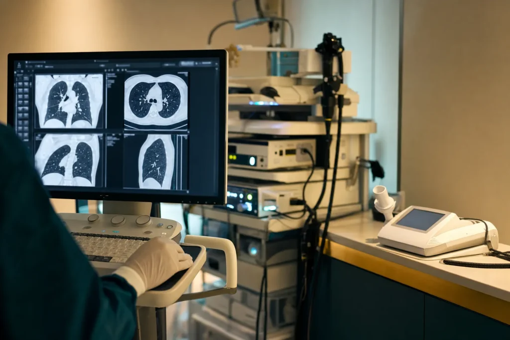
- CT scan (computed tomography): this is the key test that confirms the diagnosis. A high-resolution CT scan takes detailed cross-section images of the lungs and clearly shows widened airways, as well as how much of the lung is affected.
- Sputum sample: a sample of coughed-up phlegm is sent to the laboratory to identify which germs are present, including bacteria such as Pseudomonas aeruginosa and non-tuberculous mycobacteria. This guides the choice of antibiotics.
- Lung function tests (spirometry): you breathe into a machine to measure how much air you can move and how quickly, showing how well the lungs are working.
- Blood tests: these check for signs of infection and for underlying causes, such as problems with the immune system or certain antibody levels.
Depending on what these show, further tests may be arranged to find an underlying cause, such as a sweat test or genetic testing for cystic fibrosis, allergy tests, or a bronchoscopy (passing a thin flexible camera into the airways to look inside and take samples). There is no formal "stage 1 to 4" staging system as there is for cancer; instead your team assesses severity from the extent of damage on the scan, your lung function, how often you get flare-ups and how breathless you are.
Treatment options
Bronchiectasis cannot be cured, because the widening of the airways is permanent. However, treatment can control symptoms, reduce flare-ups and help stop the condition from getting worse. Care is usually shared by a multidisciplinary team, which may include a chest doctor (pulmonologist or respiratory physician), a specialist respiratory physiotherapist, specialist nurses, a microbiologist and sometimes a thoracic (chest) surgeon. The plan is tailored to you.
Airway clearance (chest physiotherapy). This is the cornerstone of everyday care. A respiratory physiotherapist teaches you breathing techniques and positions that help move mucus up and out, so it does not sit and breed infection. Many people use a small hand-held device that creates vibrations or back-pressure as you breathe out (an oscillating positive expiratory pressure, or PEP, device) to make clearance easier. These techniques are usually done daily, and learning to do them well makes a real difference.
Medicines. Options your team may consider include:
- Antibiotics to treat flare-ups. If you have frequent flare-ups (often defined as three or more a year), your doctor may suggest a long-term, low-dose antibiotic, sometimes a type called a macrolide, to reduce how often they happen. Inhaled antibiotics may be used for people with long-standing Pseudomonas infection.
- Mucus-thinning treatments, such as nebulised salt-water (saline) solutions breathed in as a mist, to make phlegm easier to clear.
- Bronchodilator inhalers, which relax the airway muscles to ease breathing, particularly if you also wheeze or have asthma or COPD.
- Treatment of the underlying cause, for example replacing missing antibodies in an immune problem, or treating an allergy.
Supportive care. Pulmonary rehabilitation, a supervised programme of exercise and education, is recommended if breathlessness limits what you can do, and it can improve fitness and quality of life. Vaccinations help prevent infections (see below). In severe cases, extra oxygen may be needed.
Surgery is uncommon and is mainly considered when damage is limited to one small part of the lung that keeps causing problems, or to control serious bleeding. For a small number of people with very advanced disease, a lung transplant may be discussed.
Outlook: what to expect
Bronchiectasis varies enormously from person to person, so it is not possible to predict the future for any individual, and the descriptions here are general patterns rather than a forecast for you. The single most useful message is a hopeful one: although the condition is long-term and the airway damage does not reverse, it can usually be managed well.
MedlinePlus, a service of the US National Library of Medicine, states that with treatment most people live without major disability and that the disease usually progresses slowly. The American Lung Association notes that, while there is no cure, getting the right treatment as early as possible helps people have the best possible quality of life. The NHS describes a spectrum: some people have mild symptoms that are well controlled with treatment, while in others the condition gradually worsens over time.
How things go depends on several factors, including the underlying cause, how much of the lung is affected, how often flare-ups happen, whether certain germs such as Pseudomonas are persistently present, and how consistently airway-clearance and treatment are carried out. This is encouraging, because some of these factors, particularly daily clearance, vaccinations and prompt treatment of flare-ups, are partly within your control. Regular review with a chest specialist helps keep the condition stable and catches changes early.
Living with bronchiectasis and follow-up
Many people with bronchiectasis lead full, active lives. The aim of day-to-day management is to keep the airways as clear as possible, prevent infections and stay fit. Building a steady routine usually helps the most.
- Do your airway clearance regularly, as your physiotherapist has shown you, even on good days. Consistency is what keeps mucus moving.
- Stay active. Regular exercise helps clear the lungs and keeps you fit; walking, swimming and similar activities are good choices.
- Drink enough fluids to help keep mucus thin and easier to bring up.
- Eat well and keep to a healthy weight, which supports your strength and immune system.
- Do not smoke, and avoid smoky or polluted air and other lung irritants.
- Act early on flare-ups. Learn your usual symptoms so you notice changes quickly, and know your team's plan for when phlegm increases or changes colour. Many people have an agreed "rescue" antibiotic plan.
- Wash your hands regularly to reduce the chance of picking up infections.
Follow-up is usually lifelong but need not be burdensome. Depending on severity, you may see your respiratory team once or twice a year, or more often if your condition is unstable, with repeat lung function tests and sometimes sputum checks. Living with a long-term lung condition can also affect mood; feeling low or anxious is common, and it is worth telling your team, who can offer support.
Planning treatment abroad: what affects cost and how to prepare your records
Bronchiectasis is mostly managed with ongoing medical care rather than a single operation, so planning treatment abroad usually means arranging a thorough specialist assessment, scans and tests, a personalised treatment and airway-clearance plan, and review appointments, rather than buying one fixed "procedure." Because of this, costs are highly individual and we do not list prices here. Instead, it helps to understand the factors that influence them.
Things that typically affect the cost of an assessment and treatment plan include:
- Which investigations you need, such as a high-resolution CT scan, lung function tests, sputum analysis and blood tests, and whether a bronchoscopy is required.
- The number and seniority of specialists involved, and how many appointments are needed.
- Any medicines prescribed, including nebulised or inhaled treatments.
- Whether a hospital stay is needed, for example to treat a flare-up with intravenous antibiotics.
- Whether, in the small number of suitable cases, a surgical option is being considered.
- Travel, accommodation, interpreting and follow-up arrangements.
To prepare, gather your medical records before you travel: previous chest X-rays and CT scans (ideally the actual image files on a disc or via a secure link, not only the written report), sputum and blood test results, lung function results, a list of your medicines and doses, vaccination history, and a summary of past chest infections and hospital admissions. Sharing these in advance helps the team avoid repeating tests and gives you a more accurate, personalised estimate. The best next step is to request a free consultation so your records can be reviewed and a tailored plan and estimate prepared for you.
Why Turkiye, and how to choose a good centre
Turkiye (Turkey) has become a well-established destination for international patients, with many hospitals experienced in caring for visitors from abroad and a number of facilities holding international quality accreditation. For a long-term condition like bronchiectasis, what matters most is not the destination itself but the quality and continuity of care you can arrange.
When choosing a centre, it is reasonable to verify the following:
- Accreditation. Look for recognised standards such as accreditation by Joint Commission International (JCI), which assesses hospitals against international patient-safety and quality benchmarks.
- A genuine specialist team. Bronchiectasis is best managed by a multidisciplinary team led by a chest physician (pulmonologist), with access to a respiratory physiotherapist for airway clearance, a microbiology service to identify germs and guide antibiotics, and, where relevant, a thoracic surgeon.
- Proper diagnostics on site, including high-resolution CT scanning, lung function testing and a microbiology laboratory.
- A clear follow-up plan. Because bronchiectasis needs ongoing care, ask how results, the treatment plan and any prescriptions will be shared with you and with your doctor back home, and how follow-up questions will be handled after you return.
- Support for international patients, such as interpreting, help with documents and coordination of appointments.
Avoid making decisions based on superlative marketing claims. Instead, focus on credentials you can check, the specific specialists who will see you, and how clearly the centre communicates. A reputable centre will be happy to answer these questions before you commit.
Prevention and self-care
You cannot always prevent bronchiectasis, but some steps lower the risk of developing it and, if you already have it, help protect your lungs from further damage.
- Treat chest infections promptly and complete any prescribed course of antibiotics, so infections do not linger and damage the airways.
- Keep up with vaccinations. Routine childhood vaccinations help prevent infections such as whooping cough and measles that can lead to bronchiectasis. For adults, an annual flu vaccine, the pneumococcal vaccine and COVID-19 vaccination are recommended to reduce chest infections.
- Do not smoke, and avoid second-hand smoke and other lung irritants such as fumes and heavy air pollution.
- Protect against inhaling objects, particularly in young children, and seek prompt care if something is breathed in.
If you already have bronchiectasis, the everyday self-care described earlier, regular airway clearance, staying active and hydrated, eating well, avoiding smoke and acting quickly on flare-ups, is the most powerful way to keep the condition stable. None of this replaces specialist advice. If you have a long-standing cough with phlegm or repeated chest infections, see a qualified doctor, who can arrange the right tests and, if needed, refer you to a chest specialist.
Frequently asked questions
Is bronchiectasis the same as COPD or asthma?
Can bronchiectasis be cured?
Is bronchiectasis contagious?
What is a flare-up (exacerbation) and what should I do?
Is coughing up blood serious?
How is bronchiectasis diagnosed?
Why do I need to do chest physiotherapy every day?
Do I need surgery for bronchiectasis?
Will bronchiectasis shorten my life?
Which vaccinations are recommended?
Can I still exercise and travel with bronchiectasis?
Why might someone consider having their assessment in Turkiye?
This article is for general information only and is not medical advice. Always consult a qualified doctor about your individual case.
Considering this procedure?
Send us your photos and questions. A BergemHealth coordinator and a department-head specialist will review your case and reply with honest, personalised guidance — no obligation.
Free consultation