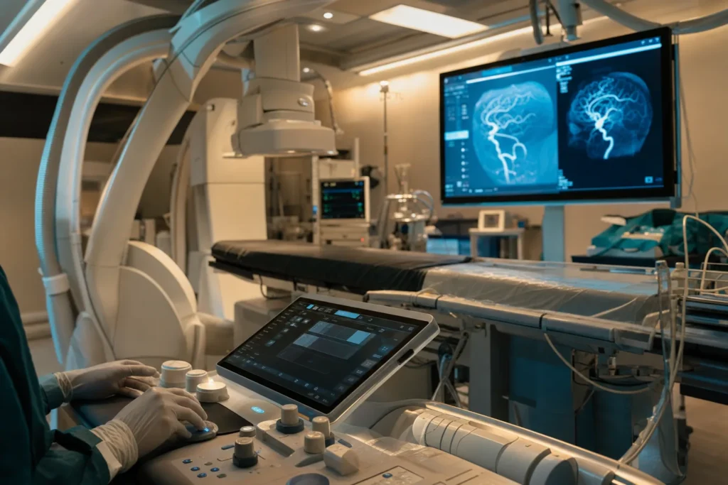
Brain aneurysm & AVM
A brain aneurysm and an arteriovenous malformation (AVM) are two different problems with the blood vessels in or around the brain. Most people who have one feel completely well and may never know it is there. This calm, plain-language guide explains what each condition is, how doctors find and treat them, what the outlook tends to be, and how to prepare your records if you are considering specialist neurosurgery in Turkiye.
What it is
This guide covers two separate conditions that affect the blood vessels of the brain. They are often discussed together because both are treated by neurosurgeons and vascular (blood-vessel) specialists, and both can, in some people, lead to bleeding inside the head. But they are not the same thing.
A brain aneurysm (also called a cerebral or intracranial aneurysm) is a weak spot on the wall of an artery in or around the brain. The constant pressure of blood pushes that weak spot outward so it balloons into a small bulge, a little like a weak area on an over-filled balloon or an old garden hose. Most aneurysms are small and cause no problems at all. The concern is that, in a minority of cases, the thin wall can leak or burst (rupture), allowing blood to escape around the brain.
An arteriovenous malformation (AVM) is a tangle of abnormal blood vessels where arteries connect directly to veins without the normal tiny vessels (capillaries) in between. Arteries carry blood at high pressure; veins are built for low pressure. In an AVM the high-pressure blood pours straight into vessels not designed to cope with it, which over time can stretch and weaken them. Most brain AVMs are present from before birth, although they are usually only discovered later in life.
The most important thing to hold onto early on is that having either condition does not mean something bad is about to happen. Many aneurysms and AVMs stay quiet for a lifetime. The purpose of medical assessment is simply to understand your individual situation and to make a calm, informed plan.
Types & subtypes
Doctors describe brain aneurysms by their shape and cause. The most common type is a saccular aneurysm, sometimes called a berry aneurysm because it looks like a small berry hanging from a stalk. These usually appear where arteries branch at the base of the brain. A fusiform aneurysm bulges out on all sides of the artery, like a spindle, rather than from one point. A mycotic aneurysm is a rarer type caused by an infection that weakens the artery wall.
Aneurysms are also grouped by size, because size is one factor in how they are managed. As a rough guide, specialists describe them as small (under about 11 mm across), large (roughly 11 to 25 mm) and giant (more than 25 mm), according to the U.S. National Institute of Neurological Disorders and Stroke (NINDS). Some people have more than one aneurysm; Cleveland Clinic notes that around 10 to 30 percent of people with an aneurysm have more than one.
AVMs vary mainly by their size, their location in the brain, and how their draining veins are arranged. The cluster of abnormal vessels at the centre is called the nidus (Latin for nest). Closely related vascular problems include dural arteriovenous fistulas (abnormal connections in the tough covering of the brain), cavernous malformations and venous malformations. These are different from a true AVM and are assessed and treated in their own ways, so it is worth asking your specialist exactly which one you have.
Causes & risk factors
For many people it is not possible to say exactly why an aneurysm formed. The NHS explains that the cause is not always clear. What doctors do understand well is the list of factors that make an aneurysm more likely to develop or to grow.
- Smoking and high blood pressure are the two factors most consistently linked to aneurysms, and both can be improved.
- Family history — having a close relative (parent, brother or sister) who has had a brain aneurysm raises your own chance.
- Inherited conditions that affect blood vessels or connective tissue, such as autosomal dominant polycystic kidney disease (ADPKD), Ehlers-Danlos syndrome, Marfan syndrome and fibromuscular dysplasia.
- Age and sex — aneurysms are most often found between ages 30 and 60, and are somewhat more common in women, according to Cleveland Clinic and NINDS.
- Cocaine and other stimulant drugs, and heavy alcohol use, which can raise blood pressure sharply.
AVMs are different. They are usually thought to form before birth as the blood vessels develop, so they are not caused by anything you did or did not do. The exact reason is still being studied, and a genetic contribution is increasingly suspected. Because they are present so early, an AVM is not something that lifestyle could have prevented.
Signs & symptoms (and when to see a doctor)
Most unruptured aneurysms cause no symptoms and are found by chance during a scan done for another reason. A larger aneurysm that presses on nearby nerves can sometimes cause warning signs such as pain above or behind one eye, a drooping eyelid, a widened (dilated) pupil, double or blurred vision, or numbness on one side of the face.
AVMs that have not bled may cause seizures (fits), persistent headaches often felt in one area, or gradual weakness, numbness or problems with speech or vision. Some people notice a rhythmic whooshing sound in the ear (pulsatile tinnitus). Many AVMs, however, cause no symptoms until they are found incidentally.
The situation that needs an emergency response is bleeding. The classic sign of a ruptured aneurysm is a sudden, extremely severe headache — often described as the worst headache of one's life, sometimes called a thunderclap headache because it peaks within seconds. It may come with a stiff neck, nausea or vomiting, sensitivity to light, double vision, confusion, seizures or loss of consciousness. A bleed from an AVM can cause similar sudden symptoms, including stroke-like weakness on one side.
Call emergency services immediately (for example 112 in Turkiye and across the EU, 911 in the United States, or your local number) for a sudden severe headache, facial drooping, sudden weakness or numbness, slurred speech, sudden vision loss or collapse. For ongoing but non-emergency symptoms such as persistent headaches, unexplained seizures or vision changes, arrange to see a doctor without delay so the cause can be investigated properly.
Screening & early detection
There is no routine screening programme of the general population for brain aneurysms or AVMs. The large majority of people will never need a brain scan to look for them, and scanning everyone would do more harm than good because it would find many tiny aneurysms that were never going to cause trouble.
Screening with a non-invasive scan such as MR angiography (MRA) or CT angiography (CTA) is sometimes offered to a small number of people at clearly higher risk — for example, those with two or more close relatives who have had a brain aneurysm, or people with certain inherited conditions such as ADPKD. Whether to be screened is a decision to make carefully with a specialist, weighing the possible reassurance against the anxiety and follow-up that finding a small aneurysm can bring.
If you think you may be in a higher-risk group, the right step is not to panic but to discuss your family and medical history with a doctor, who can advise whether imaging is sensible for you. AVMs are not screened for in this way, because they are not generally inherited in a predictable pattern.
How it is diagnosed
When a brain aneurysm or AVM is suspected — either because of symptoms or as a chance finding — doctors use detailed imaging of the blood vessels to confirm it, measure its size and map its exact position. You will not need every test; your team chooses the ones that answer the questions that matter for your care.
- CT scan: a fast scan that is excellent at showing bleeding inside the head, so it is usually the first test if a rupture is suspected.
- CT angiography (CTA): a CT scan with a contrast dye that lights up the arteries, showing the shape, size and location of an aneurysm or AVM.
- MRI and MR angiography (MRA): detailed pictures of the brain and its vessels without X-rays, useful for planning and for follow-up over time.
- Cerebral (catheter) angiography: considered the most detailed test. A thin tube is guided through a blood vessel, usually from the wrist or groin, and dye is injected to give a precise map of the vessels. It is often used when planning treatment.
- Lumbar puncture: if a scan does not show a suspected small bleed, a sample of the fluid around the spinal cord can be checked for traces of blood.
For AVMs, specialists often describe the lesion using the Spetzler-Martin grade, a simple score that adds up the size of the tangle, whether it sits in a critical (eloquent) part of the brain, and how its veins drain. A higher score generally means a more difficult and higher-risk operation, which helps the team choose the safest approach. This is a planning tool, not a personal prediction.
Treatment options
Treatment is highly individual and is decided by a multidisciplinary team — typically a neurosurgeon, an interventional (endovascular) neuroradiologist, a neurologist, anaesthetists and specialist nurses. The aim is always to weigh the risk of the condition itself against the risk of any procedure, and the choice depends on the size, shape and position of the aneurysm or AVM, your age and your overall health.
For an unruptured aneurysm, doing nothing active is often the right choice. Small aneurysms frequently never grow and have a low chance of bursting, so the NHS notes that many are simply monitored with follow-up scans to check for any change. When treatment is advised, there are two main approaches:
- Endovascular coiling: through a catheter threaded from the wrist or groin, tiny soft platinum coils (and sometimes a fine mesh tube called a stent or flow diverter) are placed inside the aneurysm so blood clots around them and the aneurysm seals off. No opening of the skull is needed.
- Surgical clipping: in an operation that opens the skull (craniotomy), the surgeon places a small metal clip across the neck of the aneurysm to stop blood entering it.
For an AVM, there are three main treatment methods, sometimes used in combination:
- Microsurgical removal: an operation to take out the tangle of vessels.
- Endovascular embolization: a catheter is used to inject a glue-like material that blocks the abnormal vessels, often to make later surgery or radiosurgery safer.
- Stereotactic radiosurgery (such as Gamma Knife): precisely focused radiation that, over months to a couple of years, causes the AVM to scar down and close. This is useful for AVMs deep inside the brain that are hard to reach.
If bleeding has occurred, treatment becomes urgent and includes both securing the aneurysm or AVM and supportive care — for example medicines such as calcium-channel blockers to reduce the artery narrowing (vasospasm) that can follow a bleed, anti-seizure medicines if needed, and sometimes a drain or shunt to relieve a build-up of fluid (hydrocephalus). Rehabilitation with physiotherapy, occupational therapy and speech therapy is often part of recovery.
Outlook / what to expect
It is important to separate two very different situations, because the outlook is not the same.
Unruptured aneurysms and AVMs that have not bled often remain stable and may never cause symptoms. The NHS points out that small aneurysms commonly do not grow and have a low risk of bursting. For an AVM, the American Stroke Association and Cleveland Clinic describe an average chance of bleeding of roughly 1 to 3 percent per year. These are population averages, not a forecast for any one person, and your own risk depends on the specific features of your lesion — which is exactly what a specialist assessment is for.
A bleed is more serious. A ruptured aneurysm is a medical emergency, and authorities including Cleveland Clinic and the American Stroke Association report that a substantial proportion of ruptures are fatal or leave lasting effects even with treatment, and that survivors may have ongoing problems with movement, speech or memory. When an AVM bleeds, sources such as the American Stroke Association and NCBI StatPearls describe a roughly 10 to 15 percent chance of death and a significant chance of permanent brain injury. These figures describe groups of patients studied over time; they cannot predict what will happen to any individual, and outcomes have generally improved as treatments and emergency care have advanced.
The clear takeaway is that the best outlook comes from finding these conditions before any bleed, getting an expert opinion, and making a calm plan together with a specialist team.
Living with it & follow-up
If your aneurysm or AVM is being watched rather than treated, follow-up usually means repeat scans at intervals your team recommends, so any change can be spotted early. Keeping these appointments is one of the most useful things you can do.
After treatment, you will normally have follow-up imaging to confirm that the aneurysm has stayed sealed or that the AVM has closed, since coiled aneurysms occasionally need a top-up procedure and radiosurgery for an AVM takes time to work. Your team will tell you when it is safe to drive, fly, return to work and resume exercise, and these timelines vary a great deal from person to person.
Day-to-day, the most helpful steps are the ones that protect your blood vessels generally: keeping blood pressure well controlled, not smoking, limiting alcohol, and avoiding cocaine and other stimulant drugs. If you take blood-thinning medication for another reason, make sure every doctor involved knows about your aneurysm or AVM. Many people also find it valuable to look after their emotional wellbeing — anxiety after a diagnosis is common and understandable, and your team or a counsellor can help. Carrying a brief note of your diagnosis and treatment, and knowing the emergency warning signs, gives both you and your family confidence.
Planning treatment abroad: what affects cost & how to prepare your records
Neurovascular care is highly individual, so there is no single price for treating a brain aneurysm or AVM. Rather than quote figures, it is more useful to understand what shapes the cost so you can ask the right questions and request a personalised estimate.
- The condition and its complexity: the size, shape and location of the aneurysm or AVM, whether there is one lesion or several, and the Spetzler-Martin grade of an AVM.
- The chosen technique: endovascular coiling or embolization, surgical clipping or removal, stereotactic radiosurgery, or a combination — each uses different equipment and theatre time.
- Implants and materials: coils, stents or flow diverters, and embolization materials.
- Hospital stay and intensity of care, including any time in intensive care and the level of monitoring needed.
- Imaging and tests before and after the procedure, and any rehabilitation.
- Whether the case is planned or an emergency, and follow-up scans afterwards.
- Practical costs of travelling such as flights, accommodation, interpreting and local transfers.
To prepare, gather your recent brain and vessel imaging (CT, CTA, MRI, MRA or catheter angiography) on disc or in digital form, any radiology reports, a summary letter from your current doctor, a list of your medicines and allergies, and notes on other health conditions. Good-quality, recent scans let a specialist team review your case accurately and give you a realistic plan. The most reliable way to understand cost is to share these records and request a personalised estimate through a free consultation.
Why Turkiye & how to choose a good centre
Turkiye has become a well-established destination for international patients seeking specialist care, including neurosurgery, with a number of hospitals that hold international quality accreditation and treat patients from around the world. As with any country, quality varies between centres, so the sensible approach is to focus on verifiable signs of a safe, experienced service rather than on marketing.
- Accreditation: look for hospitals accredited by Joint Commission International (JCI), an independent body that assesses patient safety and quality against international standards.
- A genuine multidisciplinary team: safe neurovascular care relies on neurosurgeons and interventional neuroradiologists working together, with neurology, anaesthesia, neuro-intensive care and rehabilitation on site.
- The right equipment: modern angiography suites, advanced imaging (CT and MRI), and access to both endovascular and open-surgical options so the method can be matched to your case rather than to what the centre happens to offer.
- Experience with your specific problem: ask how often the team treats aneurysms or AVMs like yours, and who exactly will perform the procedure.
- Clear communication: interpreting services, written information you understand, a named contact, and a clear plan for follow-up once you return home.
It is reasonable to ask any centre for the credentials of the treating specialists, the hospital's accreditation status, and what happens if a complication occurs. A trustworthy team will welcome these questions. A concierge service can help arrange records, second opinions, scheduling and logistics, but the medical decision should always rest with you and a qualified specialist.
Prevention, self-care & getting a second opinion
You cannot guarantee that an aneurysm will never form, and AVMs cannot be prevented at all because they develop before birth. But you can meaningfully lower the chance of an aneurysm forming or growing, and reduce the risk of a bleed, by looking after your blood vessels.
- Do not smoke — this is one of the most important and reversible factors.
- Keep blood pressure under control with regular checks, and treatment if your doctor advises it; high blood pressure is closely linked to bleeding.
- Limit alcohol and avoid cocaine and other stimulant drugs.
- Eat a balanced diet, stay active and keep a healthy weight, which all help your blood pressure and overall vascular health.
Because decisions about whether and how to treat an aneurysm or AVM involve genuine trade-offs, a second opinion is reasonable and often helpful, especially before any procedure. Getting an independent specialist to review your scans can confirm the diagnosis, clarify your options and give you confidence in the plan. There is usually time to think things through for an unruptured aneurysm or an AVM that has not bled, so do not feel rushed into a decision. The most important step of all is to have your individual situation assessed by a qualified neurovascular specialist who can explain what your particular findings mean for you.
Frequently asked questions
What is the difference between a brain aneurysm and an AVM?
Are brain aneurysms common, and will mine definitely burst?
What does a ruptured aneurysm feel like?
How is a brain aneurysm or AVM diagnosed?
What treatment options are available?
Is an unruptured aneurysm always treated?
How risky is an AVM that has not bled?
Should I be screened if a relative had a brain aneurysm?
Can a brain aneurysm or AVM be prevented?
What affects the cost of treatment, and can I get a price?
Why do people consider Turkiye for neurosurgery, and how do I choose a centre?
Is it worth getting a second opinion before treatment?
This article is for general information only and is not medical advice. Always consult a qualified doctor about your individual case.
Considering this procedure?
Send us your photos and questions. A BergemHealth coordinator and a department-head specialist will review your case and reply with honest, personalised guidance — no obligation.
Free consultation