Chronic otitis media
Chronic otitis media is a long-lasting problem of the middle ear, the small air-filled space just behind the eardrum. It usually means ongoing inflammation or repeated infection, often with a hole in the eardrum and ear discharge that keeps coming back. It can feel worrying, especially when hearing changes, but it is a well-understood condition that ear specialists treat every day with medicines and, when needed, modern ear surgery. This guide explains in plain language what the condition is, why it happens, how it is diagnosed and treated, and how to prepare if you are considering care abroad.
What chronic otitis media is
Your middle ear is a small, air-filled pocket behind the eardrum (the thin membrane called the tympanic membrane). Inside it sit three tiny bones that pass sound vibrations to the inner ear. Otitis media simply means inflammation or infection of this space. When the problem lasts a long time or keeps coming back, it is called chronic otitis media.
Doctors often describe a specific form called chronic suppurative otitis media (CSOM). "Suppurative" means pus or discharge. CSOM is defined as persistent or recurring discharge from the ear (called otorrhea) over roughly two to six weeks or longer, leaking out through a hole (perforation) in the eardrum that has not healed.
This is different from a sudden, short-lived ear infection (acute otitis media), which often clears on its own within a few days. Chronic otitis media is the long-running version, where the eardrum may have a lasting hole, the lining stays inflamed, and discharge or hearing changes return again and again.
It is a common condition worldwide. Estimates suggest that chronic suppurative otitis media affects somewhere between 65 and 330 million people globally, and it is one of the most frequent causes of preventable hearing loss, especially in lower-income communities.
Types and subtypes
Ear specialists (called otolaryngologists or ENT doctors) divide chronic otitis media into a few patterns. Knowing which type you have shapes the treatment.
- Chronic otitis media with effusion ("glue ear"). Here the middle ear fills with sticky fluid without active infection. There is usually no hole in the eardrum and no discharge, but hearing can be muffled. It is most common in children.
- Chronic suppurative otitis media (CSOM), mucosal type. There is a lasting hole in the eardrum and recurring discharge, but the disease stays in the lining (mucosa) of the middle ear. This is sometimes called the "safe" or active mucosal form.
- Chronic otitis media with cholesteatoma (squamous type). A cholesteatoma is an abnormal but non-cancerous build-up of skin cells that forms a growing pocket or cyst in the middle ear. Over time it can erode the tiny ear bones and surrounding structures, so this type needs closer attention.
Doctors also describe disease as active (currently discharging or growing) or inactive (a dry hole or healed pocket with no current infection). These labels simply help the team decide whether you need treatment now or watchful monitoring.
Causes and risk factors
Chronic otitis media usually develops after repeated or poorly resolved ear infections, often starting in early childhood. A central reason is trouble with the Eustachian tube — the narrow channel that connects the middle ear to the back of the nose and keeps air pressure balanced and the space ventilated. When this tube does not work well, fluid and bacteria can build up, the eardrum can become damaged, and inflammation can persist.
Common contributing factors include:
- Frequent episodes of acute (short-term) middle ear infection in childhood.
- Repeated colds and upper respiratory infections.
- A hole in the eardrum from infection, injury, or a previous grommet (ear tube).
- Eustachian tube dysfunction, which is found in a large share of people who eventually need ear surgery.
- Crowded living conditions, exposure to tobacco smoke, and poorer nutrition or hygiene, which raise risk at a population level.
- Certain anatomical conditions, such as cleft palate or Down syndrome, which affect how the ear and Eustachian tube work.
The bacteria most often involved in active discharge include Pseudomonas aeruginosa and Staphylococcus aureus. Having these risk factors does not mean you have done anything wrong — many people develop chronic ear problems simply from the way their ears and Eustachian tubes are built.
Signs and symptoms (and when to see a doctor)
Symptoms vary from very mild to more troublesome. The two most common are:
- Ear discharge (otorrhea). This is often the leading sign. It may be persistent or come and go, and it can sometimes be foul-smelling. It is frequently painless.
- Hearing loss. Usually this is conductive hearing loss, meaning sound is not passing efficiently through the damaged eardrum and ear bones. It often sounds muffled rather than completely absent.
Other possible symptoms include a feeling of fullness or pressure in the ear, ringing in the ear (tinnitus), and sometimes a dull ache.
It is wise to see a doctor if you have ear discharge that lasts more than a few days, hearing that is getting worse, or repeated ear infections. Seek prompt medical attention if you notice any of these warning signs, which can point to a cholesteatoma or a spreading infection: dizziness or a spinning sensation (vertigo), severe or new ear pain, fever, weakness of the face on the affected side, severe headache, or sudden marked hearing loss. These are uncommon, but they are worth acting on quickly.
Screening and early detection
There is no national screening programme that tests healthy adults specifically for chronic otitis media. Because the condition almost always announces itself with discharge or hearing changes, the practical path to early detection is simple: take ongoing ear symptoms seriously and have your ears examined.
Early detection mostly happens through everyday care:
- Newborn and childhood hearing checks. Routine hearing screening in babies and school-age children can flag the kind of hearing loss that sometimes signals ongoing middle-ear disease, prompting an ENT review.
- Acting on persistent discharge. Ear discharge that does not settle, or that returns repeatedly, should be checked by a clinician rather than waited out.
- Following up after acute infections. If an ear infection does not fully clear, a follow-up examination helps catch a problem before it becomes chronic.
Catching a cholesteatoma early matters most, because removing it before it damages the ear bones or inner ear gives the best chance of preserving hearing and avoiding complications.
How it is diagnosed
Diagnosis is usually straightforward and starts with a careful look inside the ear. The main steps include:
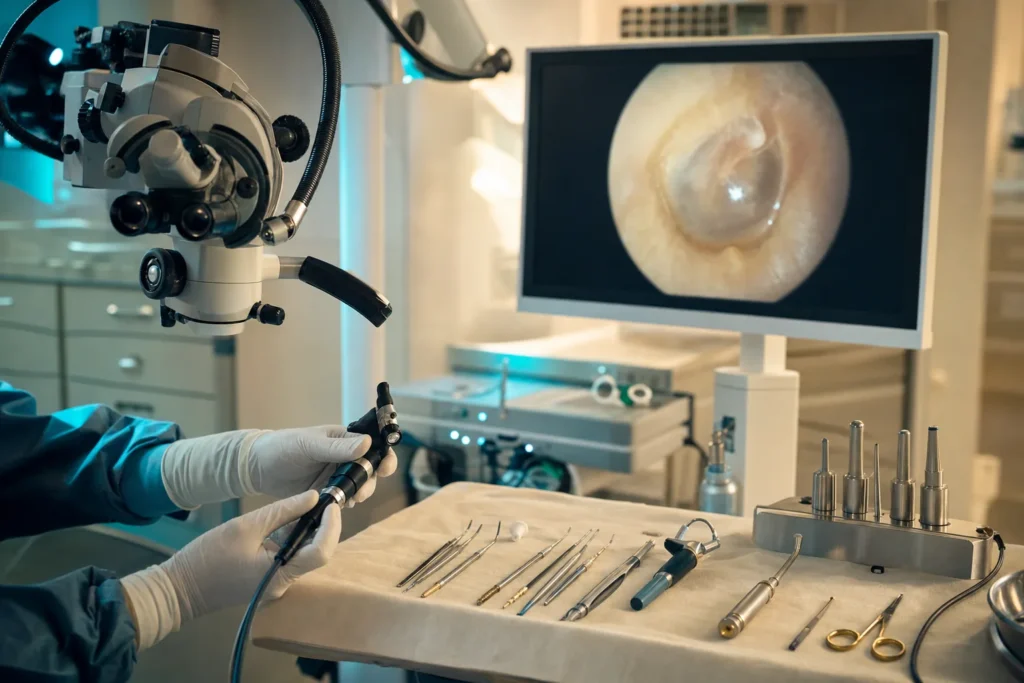
- Otoscopy. The doctor uses a lighted instrument (an otoscope, sometimes a microscope or endoscope) to inspect the eardrum, looking for a perforation, discharge, an inflamed lining, or the whitish debris of a cholesteatoma.
- Hearing tests (audiometry). An audiologist measures how well you hear different sounds. This shows the type and degree of any hearing loss and gives a baseline before treatment.
- Tympanometry. A painless test that checks how the eardrum moves and whether there is fluid behind it.
- Swab and culture. If the ear is discharging, a small sample may be taken to identify the bacteria and guide the right antibiotic.
- Imaging. A CT scan is often used when a cholesteatoma is suspected, to map the extent of disease and check the surrounding bone. An MRI may be added in selected cases.
There is no formal cancer-style "staging" for this condition. Instead, the team describes whether disease is active or inactive, whether a cholesteatoma is present, and how much the eardrum, ear bones, and hearing are affected. This picture guides whether medicine alone is enough or whether surgery is advisable.
Treatment options
Treatment is tailored to your type of disease, your hearing, and whether a cholesteatoma is present. Care is usually delivered by a multidisciplinary team — an ENT surgeon, an audiologist, nurses, and sometimes specialists in imaging and infection.
Medical (non-surgical) treatment
- Ear cleaning (aural toilet). Gently clearing discharge and debris, often by a clinician, helps medicines reach the middle ear and lets the doctor see clearly.
- Topical antibiotic ear drops. These are the mainstay for an actively discharging ear. Quinolone-type drops are widely recommended because they are effective and avoid the small risk of inner-ear harm linked to some older drops.
- Oral or intravenous antibiotics. Reserved for more severe or resistant cases.
- Keeping the ear dry. Protecting the ear from water during washing or swimming helps prevent flare-ups while a perforation is present.
Surgical treatment
Surgery is considered when medicine does not control the disease, when there is a lasting eardrum hole, when hearing needs restoring, or when a cholesteatoma is present (where surgery is generally the only effective treatment). Common operations include:
- Tympanoplasty. Repair of the eardrum perforation, sometimes with reconstruction of the tiny hearing bones.
- Mastoidectomy. Removal of infected or diseased tissue from the mastoid bone behind the ear, often combined with tympanoplasty, especially for cholesteatoma.
Surgeons choose between techniques (for example, keeping the ear canal wall intact versus removing it) based on how widespread the disease is. For some cholesteatomas, a planned second operation is done later to check that none has come back.
Supportive care
If hearing loss remains after treatment, hearing aids or other devices can help. Audiology support, regular ear care, and clear advice on keeping the ear dry are all part of good long-term management.
Outlook: what to expect
The outlook for chronic otitis media is generally good when it is properly treated and complications are avoided. Many people with the mucosal (non-cholesteatoma) form do well with ear drops, ear cleaning, and, where appropriate, surgery to repair the eardrum and improve hearing.
It is honest to say that results vary from person to person. Hearing improvement after eardrum or ear-bone surgery depends on how much damage was already present, and some hearing loss can remain. Cholesteatoma can sometimes return after surgery, which is why follow-up examinations are important.
The main concern with untreated chronic disease, particularly cholesteatoma, is the slow risk of complications as it spreads. These can include further hearing loss, balance problems, facial weakness, and, rarely, infection spreading toward the brain. The encouraging point is that timely diagnosis and modern surgery are designed precisely to prevent these outcomes. None of these figures or risks is a prediction for any individual — your own outlook depends on your specific ear, and your ENT specialist is the right person to discuss it with you.
Living with it and follow-up
Living well with chronic otitis media is mostly about steady, sensible ear care and keeping in touch with your specialist.
- Keep the ear dry when advised. If you have a perforation or have had surgery, your team may suggest avoiding getting water in the ear — for example, using a petroleum-jelly-coated cotton plug when bathing and avoiding swimming until cleared.
- Treat flare-ups early. Start prescribed ear drops promptly when discharge returns, and have the ear cleaned if your clinician recommends it.
- Attend follow-up appointments. After cholesteatoma surgery in particular, regular checks (often over the following year and sometimes longer) are used to catch any recurrence early.
- Support your hearing. If hearing loss affects daily life, ask about hearing aids and practical strategies. Untreated hearing loss in children can affect speech and learning, so children deserve prompt attention.
- Manage colds and allergies. Looking after upper-airway health can reduce the Eustachian tube problems that fuel ear disease.
Many people manage the condition for years with minimal disruption once a clear plan is in place.
Planning treatment abroad: what affects cost and how to prepare your records
If you are considering having ear surgery in another country, the most useful first step is to gather your medical information so a specialist can give you an accurate, personalised plan. Rather than quoting prices here, it helps to understand the factors that influence the overall cost and complexity of care:
- Which operation you need. A simple eardrum repair (tympanoplasty) is different in scope from a mastoidectomy or cholesteatoma removal with hearing-bone reconstruction.
- Whether a cholesteatoma is present and how far it has spread, which affects surgical time and whether a second-stage procedure is planned.
- One ear or both.
- The tests required before surgery, such as audiometry and CT imaging.
- Length of hospital stay and anaesthesia needs, which the surgical team determine for your case.
- Follow-up arrangements and any rehabilitation, such as hearing aids.
To prepare, collect your recent hearing test results, any previous ear scans (CT or MRI) on disc or in digital form, a summary of past ear infections and surgeries, and a current medication list. Sharing these allows a centre to review your case remotely and arrange a free consultation and a tailored estimate before you travel. Always request a written, itemised quote that lists what is and is not included.
Why Turkiye and how to choose a good centre
Turkiye (Turkey) has become a well-established destination for ear, nose, and throat surgery, with experienced ENT surgeons, modern hospitals, and international patient services that help with travel, translation, and scheduling. As with anywhere, the key is choosing the centre carefully rather than by reputation alone.
When evaluating a hospital or surgeon, it is reasonable to verify:
- Accreditation. Look for recognised quality accreditation, such as Joint Commission International (JCI), and proper national licensing of the hospital.
- Surgeon credentials and experience specifically in ear (otologic) surgery, including tympanoplasty and cholesteatoma surgery.
- A genuine multidisciplinary team, including audiology and access to good imaging.
- Clear information about the proposed procedure, the technique, expected results, risks, and what follow-up is offered if you return home.
- Transparent, written pricing and a realistic plan for managing complications or revision surgery.
A trustworthy centre will welcome your questions, give honest answers about what surgery can and cannot achieve for your hearing, and avoid pressure or sweeping promises. A second opinion is always reasonable before agreeing to an operation.
Prevention and self-care
Not every case can be prevented, but several steps lower the risk of chronic ear disease and flare-ups:
- Treat acute ear infections properly and have them followed up if they do not fully clear.
- Keep vaccinations up to date. Vaccines that reduce common childhood infections, including pneumococcal vaccination, have helped lower rates of acute otitis media that can lead to chronic disease.
- Avoid tobacco smoke, including secondhand smoke around children.
- Protect the ear from water if you have a perforation, and avoid pushing cotton buds or objects into the ear canal.
- Look after general health — good nutrition and managing colds and allergies support healthy Eustachian tube function.
If you already have chronic otitis media, the best "self-care" is partnership with your ENT team: treating discharge early, keeping follow-up appointments, and asking about hearing support when you need it. Population-level improvements in housing, hygiene, and nutrition have been shown to substantially reduce how common this condition is, underlining how much everyday factors matter.
Frequently asked questions
What is the difference between chronic otitis media and a normal ear infection?
Is chronic otitis media dangerous?
Will my hearing come back after treatment?
What is a cholesteatoma and why does it matter?
Do I always need surgery for chronic otitis media?
Can chronic otitis media come back after surgery?
Why is keeping the ear dry so important?
Is ear surgery for this condition painful, and how long is recovery?
How do I prepare to get a treatment plan from a hospital abroad?
What should I check before choosing a hospital in Turkiye?
This article is for general information only and is not medical advice. Always consult a qualified doctor about your individual case.
Considering this procedure?
Send us your photos and questions. A BergemHealth coordinator and a department-head specialist will review your case and reply with honest, personalised guidance — no obligation.
Free consultation