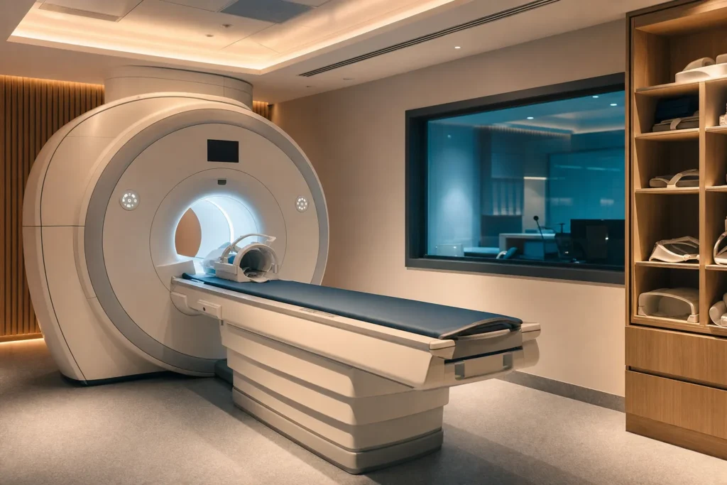
3T MRI scan
An MRI scan lets doctors look inside your body in remarkable detail without any radiation, using a strong magnet and radio waves instead of X-rays. A "3T" (3 Tesla) scanner is a more powerful version that produces sharper, more detailed pictures, often faster. This guide explains what a 3T MRI scan is, who it helps, how it feels on the day, what it can and cannot do, the realistic risks, indicative costs in Turkiye (Turkey), and how to choose a safe, properly accredited clinic.
- Anaesthesia
- None for most scans; mild sedation or, rarely, general anaesthesia for severe claustrophobia or young children.
- Duration
- About 15 to 60 minutes per body area; some detailed or multi-region scans take up to 90 minutes.
- Recovery
- Immediate for a plain scan; you can resume normal activity at once. If sedated, allow a day and do not drive for 24 hours.
- Hospital stay
- None. MRI is an outpatient test; you go home the same day.
Indicative hospital range, confirmed after your file is reviewed. You pay the hospital’s price — we add no markup.
What a 3T MRI scan is
MRI stands for magnetic resonance imaging. It is a painless test that creates detailed pictures of the inside of your body using a very strong magnet, radio waves and a computer. Crucially, it uses no X-rays and no radiation, which is one reason doctors rely on it so heavily.
Here is the idea in plain terms. Your body is mostly water, and water contains hydrogen atoms. Inside the scanner, the powerful magnet briefly lines up the tiny hydrogen particles in your tissues. A short pulse of radio energy nudges them out of line; when the pulse stops, they spring back and give off a faint signal. A receiving coil picks up those signals, and a computer turns them into cross-sectional images, like thin slices through the body that can be viewed from many angles.
The "3T" part refers to the strength of the magnet. Tesla (T) is the unit used to measure magnetic field strength. A 1.5T scanner, the long-standing workhorse, is already roughly 30,000 times stronger than the Earth's magnetic field. A 3T (3 Tesla) scanner is about twice as powerful again. That extra strength produces a stronger signal from your tissues, which generally means higher-resolution images and, often, a faster scan. MRI is especially good at showing soft tissues, organs, the brain and spinal cord, joints, muscles and blood vessels, areas where X-ray and CT scans are less informative.
Who a 3T MRI scan is for, and who should avoid it
An MRI may be requested for a huge range of reasons. Common ones include investigating problems in the brain and spinal cord (such as suspected stroke, multiple sclerosis, tumours, aneurysms or persistent headaches), the joints and muscles (knee, shoulder, hip and back injuries, cartilage and ligament tears), the abdomen and pelvis (liver, kidneys, pancreas, bowel, bladder and reproductive organs), the prostate, the breast (alongside mammography), the heart and blood vessels, and to monitor a known condition or check how well a treatment is working.
A 3T scanner is particularly valued where fine detail matters, for example small structures in the brain, the pituitary gland and cranial nerves, prostate imaging, and detailed joint imaging in sports and orthopaedic injuries. For some uses, though, a 1.5T scanner is preferred, for instance certain lung studies or scanning around some metal implants, where the higher field can create more image distortion.
Some people should not have an MRI, or need careful checking first. You may be unable to have a standard scan, or need special arrangements, if you have:
- A cardiac pacemaker or defibrillator that is not certified MRI-safe, or other older implanted electronic devices.
- Certain aneurysm clips in the brain, a cochlear (inner-ear) implant, or some nerve stimulators.
- Metal fragments in or near the eyes, or bullets or shrapnel in the body.
Many modern implants, including most newer pacemakers, joint replacements and stents, are labelled MRI-conditional, meaning they can be scanned safely under specific conditions. The clinic needs the exact device and manufacturer details to confirm this. Tell staff about any surgery, implant, tattoo or permanent make-up, kidney problems, allergies, and whether you are or might be pregnant.
Types and techniques
"MRI scan" is really an umbrella term. The scanner can be set up in many ways depending on what your doctor needs to see:
- Standard (anatomical) MRI, the everyday scan of the brain, spine, joints or organs.
- MRI with contrast, where a dye called gadolinium is given through a vein to make blood vessels, inflammation and some tumours show up more clearly.
- MR angiography (MRA), which maps blood vessels, often without any dye.
- Functional MRI (fMRI), which detects tiny changes in blood flow to show which parts of the brain are active during a task, used in planning brain surgery and in research.
- Multiparametric MRI, which combines several sequences in one sitting, widely used for the prostate.
- Cardiac MRI, which images the beating heart, its chambers and blood flow.
There is also a choice of scanner design. A traditional "closed-bore" machine is a cylinder, like a large doughnut with a tunnel, and gives the best image quality. An "open" or wide-bore MRI has more space around you, which helps people who feel anxious in enclosed spaces or who are larger, though open scanners often use a lower field strength and may produce slightly less detailed images. Modern 3T systems increasingly come in wider, shorter-bore designs that balance comfort with image quality.
How a 3T MRI is done: anaesthesia, steps and timing
An MRI is an outpatient test and almost always done while you are fully awake. No anaesthesia is needed for most people. If you find enclosed spaces very difficult, the clinic can offer mild sedation; this is needed by roughly 1 in 20 patients. General anaesthesia is reserved for unusual cases, such as young children or people who genuinely cannot lie still.
A typical visit goes like this:
- Safety screening. You complete a questionnaire about implants, metal, surgery, allergies and pregnancy, then change into a gown and remove all metal items, jewellery, watches, hearing aids, hairpins, coins and anything magnetic. This step protects you, because loose metal can be pulled violently toward the magnet.
- Contrast, if needed. If your scan uses gadolinium dye, a small drip line is placed in your arm. You may have a blood test beforehand to check your kidney function.
- Positioning. You lie on a padded couch that slides into the tunnel. The body part being scanned is placed at the centre of the magnet. You are given earplugs or headphones, because the scanner makes loud tapping and knocking noises while it works.
- The scan. You must stay as still as possible while a series of image sequences runs. The radiographer watches from an adjacent room and talks to you through an intercom; you hold a buzzer to call for attention. It is normal for the scanned area to feel slightly warm.
The imaging itself usually takes about 15 to 60 minutes per body region. Detailed or multi-region studies can take up to around 90 minutes. If contrast was used, you may be watched for about 30 minutes afterward as a precaution.
Recovery, step by step
For a standard MRI without sedation, there is effectively no recovery period. You can get dressed, eat, drive and return to work or normal activity straight away. The test does not leave any after-effects in your body, because nothing radioactive is involved.
If your scan used gadolinium contrast, you will simply be encouraged to drink plenty of fluids over the next day to help your kidneys clear the dye. Mild, short-lived effects such as a metallic taste, a brief headache or slight nausea occasionally occur and settle on their own. If you are breastfeeding, current guidance generally considers it safe to continue; ask staff if you have any concern.
If you were given sedation, plan to take it easy for the rest of the day. You should not drive for 24 hours, and you will need a responsible adult to take you home and ideally stay with you until the medication fully wears off.
Risks and possible complications
For the average patient, an MRI carries almost no risk when staff follow proper safety procedures. There is no radiation. The main hazards are mechanical and relate to the magnet, which is why the safety screening is taken so seriously:
- The projectile effect. The magnet is always on and can turn loose metal objects into dangerous projectiles. This is why all metal must be removed before you enter the room.
- Implant and device problems. An incompatible implant could heat up, move or stop working. Correct screening prevents this, which is why your device details matter so much.
- Contrast reactions. Gadolinium rarely causes allergic reactions, and far less often than the iodine dyes used in CT. In people with serious kidney disease, older types of gadolinium were linked to a rare but serious condition called nephrogenic systemic fibrosis (NSF), which thickens the skin and tissues. With the newer agents used today and proper kidney checks, this risk is now extremely low. Small traces of gadolinium can remain in the body for a time; this is monitored by regulators, and contrast is only used when it adds clear value.
- Tattoos and permanent make-up. A small minority of older inks contain iron oxides that can tingle, feel warm or, very rarely, cause a minor skin burn, and can blur the image locally. Reactions are uncommon and usually mild; tell the radiographer about tattoos and report any heat or discomfort at once.
- Claustrophobia. Feeling anxious in the tunnel is common but not dangerous, and can be managed with reassurance, an open scanner or mild sedation.
Pregnancy: there is no known risk to having an MRI while pregnant, but non-urgent scans are often postponed, and contrast dye is usually avoided as a precaution unless it is essential.
Results and how long they last
An MRI is a diagnostic test, not a treatment, so "results" here means the images and the report, not a lasting physical change. You will not normally be told the findings on the day. A radiologist, a doctor specially trained to interpret scans, studies the images and writes a detailed report.
That report is sent to the doctor who referred you, who then explains what it means and what happens next. Turnaround varies by clinic but is typically within a few days to about 1 to 2 weeks; many private and medical-tourism clinics provide a written report and a copy of the images on a disc or via a secure online link, often more quickly.
The images are a snapshot of your body at that moment. If you are being monitored for a condition, your doctor may compare a future scan with this one, which is why keeping your images and report is useful. A 3T scan's higher resolution can sometimes pick up small findings that a lower-field scan might miss, which is helpful for accuracy but occasionally leads to extra tests to clarify a minor, harmless finding.
Costs: indicative ranges and what changes the price
MRI pricing depends heavily on the country, the clinic, the body area and whether contrast is used. In Turkiye, a single-region 3T MRI at a private clinic commonly falls in the region of EUR 150 to EUR 700, with straightforward scans (such as a single joint or the brain without contrast) at the lower end and more complex studies (contrast-enhanced, multi-region, prostate multiparametric or whole-body screening) toward the higher end. These figures are indicative ranges only, not a quote; the actual price varies by case, the referring or supervising doctor, and the individual clinic.
Factors that move the price include:
- How many body regions are scanned (each area is usually priced separately).
- Whether gadolinium contrast is used, which adds the cost of the dye and the placement of a drip line.
- The type of study, with specialised scans such as cardiac MRI, MR angiography, functional MRI or multiparametric prostate MRI costing more than a standard scan.
- Scanner and clinic, including 3T versus 1.5T, accreditation level and location.
- Extras such as sedation, a written radiologist report in your language, a second opinion, and any package that bundles a consultation or imaging disc.
Compared with the United States, the United Kingdom and much of Western Europe, MRI in Turkiye is often substantially less expensive. Always ask for an itemised, written estimate that states exactly which regions are included and whether contrast and the report are part of the price.
Why people travel to Turkiye, and how to choose a safe clinic
Turkiye has become a major hub for medical imaging and treatment because it combines modern, well-equipped hospitals, including many with 3T scanners, with prices well below those in many Western countries, and short waiting times. The country also has a large number of internationally accredited facilities.
To choose a safe clinic, focus on verifiable credentials rather than marketing claims:
- Accreditation. Look for hospitals accredited by Joint Commission International (JCI), a globally recognised standard for patient safety and quality. You can confirm a facility's status on JCI's own "find accredited organizations" directory. Match the exact hospital name and address to the place where your scan will actually be done.
- Who reads your scan. Ask whether a board-certified radiologist will interpret the images, and whether you will receive a full written report (ideally in your language) plus the raw images.
- The equipment. Confirm the field strength (1.5T or 3T) and that the scanner suits your needs. For implants, confirm the clinic can check MRI-conditional device labelling.
- Transparency. A trustworthy clinic gives clear, itemised written pricing, explains what is and is not included, and answers questions about safety screening and contrast.
Get your imaging and report in a portable format so your own doctor at home can review it. If a scan is being used to plan treatment, make sure the imaging protocol matches what your treating team will need.
How to prepare and what to ask at your consultation
Preparation for most MRIs is simple. For many scans you can eat, drink and take your usual medicines as normal; some abdominal or pelvic studies require a few hours of fasting, and the clinic will tell you in advance. Wear comfortable clothing without metal fasteners, and leave valuables and metal jewellery at home or be ready to remove them. Arrive with a list of your implants, past surgeries, allergies and current medications.
Tell the clinic beforehand if you:
- Have any implant or device, metal fragments, or work that exposed you to metal (the exact device model helps).
- Have kidney problems, diabetes, or have ever reacted to contrast dye.
- Are pregnant, might be pregnant, or are breastfeeding.
- Have tattoos or permanent make-up, or struggle with enclosed spaces.
Useful questions to ask include: Which body areas will be scanned, and will contrast be used? Is the scanner 1.5T or 3T? Who interprets the images, and when and how will I get my report? Is the price itemised and final? Will I receive a copy of the images? What sedation options are available if I feel claustrophobic, and will I then need someone to take me home? If you are travelling, also ask how the results will be shared with your home doctor.
Aftercare and travelling for treatment, including when it is safe to fly
Because an MRI is non-invasive, aftercare is minimal. After a plain scan you can carry on with your day immediately. After a contrast scan, drink plenty of fluids to help clear the dye, and watch for any delayed reaction such as a rash or itching, which is uncommon. If you had sedation, rest, avoid driving for 24 hours and have someone accompany you.
For people travelling to Turkiye, the good news is that there is no specific medical reason that an MRI itself prevents you from flying. There is no radiation to clear and no recovery from the imaging. The usual caveats apply: if you received sedation, do not fly until it has fully worn off and you feel completely normal, ideally with clearance from the clinic. If you had contrast, stay well hydrated and be aware of the small chance of a delayed allergic reaction; if you are anxious about this, ask the clinic before you travel.
Practical travel tips: schedule the scan with enough buffer before any return flight so you can attend a follow-up consultation and collect your written report and imaging disc. Carry digital and printed copies of the report and images so your doctor at home can review them. If the MRI is part of a wider treatment plan, coordinate the timing so the images are available before any procedure, and keep the clinic's contact details handy in case you have questions once you are home.
Frequently asked questions
Does an MRI use radiation like an X-ray or CT scan?
What is the difference between a 3T and a 1.5T MRI?
Is an MRI scan painful?
How long does an MRI take?
Can I have an MRI if I have a pacemaker or metal implant?
What is contrast dye, and is gadolinium safe?
I am claustrophobic. What are my options?
Are tattoos a problem for an MRI?
When will I get my MRI results?
Is it safe to fly after an MRI?
How much does a 3T MRI cost in Turkiye?
How do I check a Turkish clinic is safe and accredited?
This article is for general information only and is not medical advice. Always consult a qualified doctor about your individual case.
Considering this procedure?
Send us your photos and questions. A BergemHealth coordinator and a department-head specialist will review your case and reply with honest, personalised guidance — no obligation.
Free consultation