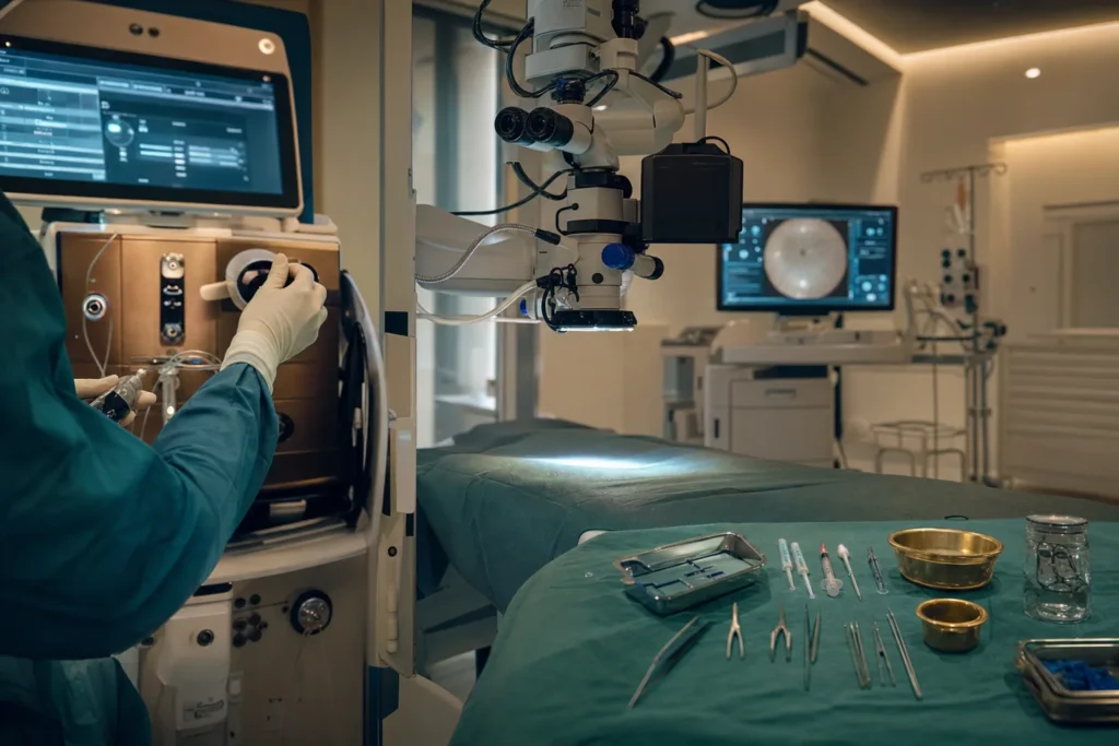
Vitreoretinal surgery
Vitreoretinal surgery is a group of delicate operations on the back of the eye that can save sight from conditions like retinal detachment, diabetic bleeding and macular holes. This guide explains, in everyday language, what the surgery involves, how recovery works, and what to check before you travel for treatment.
- Anaesthesia
- Usually local anaesthetic with sedation; general anaesthesia for children or anxious patients
- Duration
- About 1 hour for simple cases, up to 2-3 hours for complex ones
- Recovery
- Drops for several weeks; often 2-4 weeks off work; vision settles over weeks to months
- Hospital stay
- Usually a day case; home within 1-2 hours, occasionally one overnight stay
Minimum price, for reference only — the final cost is calculated individually after your records are reviewed. You pay the hospital’s price; our service is free for you.
What vitreoretinal surgery is
Vitreoretinal surgery is the branch of eye surgery that treats problems at the back of the eye, where the retina and the vitreous sit. The retina is the thin, light-sensitive layer that lines the inside back wall of your eye, a bit like the film in an old camera. It captures light and sends pictures to your brain. The vitreous (say it "VIT-ree-us") is the clear, jelly-like substance that fills the middle of the eyeball and helps it keep its round shape.
The most common vitreoretinal operation is a vitrectomy ("vit-RECK-toe-mee"), which simply means removing the vitreous gel. Surgeons do this when the gel is cloudy, is pulling on the retina, or is in the way of the repair they need to make. Once the gel is removed, the surgeon can work directly on the retina, and then fills the space with a temporary substitute, sterile salt water (saline), a gas bubble, or silicone oil, depending on what the eye needs.
This is a routine, well-established operation. In the United States alone, surgeons perform roughly 225,000 vitrectomies every year. It is usually carried out by a retina specialist, an ophthalmologist (eye doctor) who has done extra training in this specific area.
Who is a good candidate
Vitreoretinal surgery is not a lifestyle or cosmetic choice; it is recommended when a specific problem at the back of the eye threatens your sight. Common reasons include:
- Retinal detachment, when the retina lifts away from the wall of the eye. This is a medical emergency because untreated detachment usually leads to permanent vision loss in that eye.
- Diabetic retinopathy, where diabetes damages the tiny blood vessels in the retina. In advanced cases these fragile vessels bleed into the vitreous (a vitreous haemorrhage) or form scar tissue that pulls the retina off.
- Macular hole, a small gap in the macula (the central part of the retina you use for reading and recognising faces).
- Macular pucker (also called an epiretinal membrane), a thin layer of scar-like tissue that wrinkles the retina and distorts vision.
- Severe eye infection (endophthalmitis), serious injury, or complications from earlier cataract surgery.
- Sometimes persistent, vision-blocking floaters when other options have failed.
Who should be cautious? Surgery may be delayed or reconsidered if you have an active eye infection in the surface tissues, are unwell from another condition that makes anaesthesia risky, or cannot follow the important after-care, especially the face-down positioning some cases require. People who cannot avoid flying for several weeks may not be suitable for the gas-bubble approach. Your surgeon weighs the risk of operating against the risk of leaving the problem untreated, and for sight-threatening conditions the balance usually favours treatment.
Types and techniques
There are a few different vitreoretinal techniques, and your surgeon may combine them in one sitting.
Pars plana vitrectomy is the standard approach for problems at the back of the eye. The surgeon enters through the pars plana, a safe zone in the white of the eye (the sclera) that avoids the retina and the lens. Modern vitrectomy uses tiny, self-sealing incisions only about half a millimetre wide, which often need no stitches. This "small-gauge" surgery (you may see the terms 23-, 25- or 27-gauge) is generally more comfortable and allows faster visual recovery than older methods.
Anterior vitrectomy deals with vitreous gel that has moved into the front part of the eye, usually after trauma or a complication during cataract surgery.
To hold the retina in place while it heals, the surgeon may add a tamponade (a temporary internal support):
- Gas or air bubble, which the body absorbs on its own over days to weeks.
- Silicone oil, used for more complex cases; it stays longer and is usually removed in a second, smaller operation later.
Vitrectomy is also frequently combined with other steps in the same procedure, such as laser treatment (endolaser) to seal retinal tears, peeling away scar membranes with fine forceps, or a scleral buckle (a soft silicone band placed around the outside of the eye) to support a detached retina.
How it is done
Anaesthesia. Most vitrectomies are done under local anaesthetic, which numbs and freezes the eye while you stay awake but very comfortable, often with sedation ("twilight" medicine) to help you relax. You will not see the operation or feel pain. General anaesthesia (fully asleep) is used less often, mainly for children, very anxious patients, or particularly complex cases.
The steps. After your eye is numbed and the pupil widened with drops, the surgeon makes two or three tiny openings in the white of the eye. Through these they pass fine instruments and a thin light. A device called a vitrector gently cuts and removes the vitreous gel. The surgeon then carries out the specific repair, for example peeling scar tissue, sealing a tear with laser, or flattening a detached retina. Finally the space is filled with saline, a gas bubble, or silicone oil. The small incisions usually seal themselves without stitches.
How long it takes. A straightforward vitrectomy typically takes about one hour. More complex repairs, such as an advanced retinal detachment or heavy diabetic scarring, can take two to three hours or more.
Recovery, step by step
Straight after surgery. Vitrectomy is usually a day case. After local anaesthetic many people are ready to go home within about an hour; after general anaesthetic you typically stay a little longer, at least a couple of hours, until you are fully awake. Your eye will be covered with a pad or shield, and you will be given eye drops and instructions.
The first days. A gritty or scratchy feeling, as if something is in the eye, is common and normal. Mild swelling or bruising of the eyelid may appear. You will use antibiotic and anti-inflammatory (steroid) drops for several weeks to prevent infection and calm inflammation.
Positioning. If a gas bubble or silicone oil was used, you may need to hold a particular head position, often face-down, so the bubble presses on the right part of the retina. A common instruction is to maintain the position for about 50 minutes of every hour for a few days. Your team will tell you exactly what to do, as it depends on your case.
Your vision. Expect blurry vision at first. While a gas bubble is present, sight in that eye is poor and you may see the bubble as a dark line that gradually shrinks. Air bubbles clear in about 7 to 10 days; gas bubbles take roughly 2, 4 or 8 weeks depending on the type used. Final vision is usually only assessed once the bubble has gone, often around six weeks.
Back to normal. Many people take about two to four weeks off work, longer for heavy or dusty jobs. You should not drive until your eye team confirms it is safe, and you must avoid air travel while a gas bubble is in the eye.
Risks and possible complications
Vitreoretinal surgery is generally safe, and serious complications are rare, but no eye operation is risk-free. Your surgeon should explain the specific risks for your situation. Possible problems include:
- Cataract, a clouding of the eye's natural lens. This is the most common longer-term effect and is especially likely in people over 50. It can be corrected with a separate, routine cataract operation.
- Raised eye pressure, which is fairly common after surgery (reported in roughly 1 in 5 to 1 in 10 cases) and is usually managed with extra drops.
- Infection inside the eye (endophthalmitis), which is rare, around 1 in 1,000 operations, but serious if it occurs.
- A new retinal tear or detachment, reported in fewer than 2 in 100 patients, sometimes needing a further operation.
- Bleeding inside the eye.
- Severe loss of vision, which is uncommon, on the order of 1 in 1,000.
Report any increasing pain, sudden drop in vision, growing redness or discharge to your surgical team straight away, as prompt treatment matters.
Results and how long they last
For many conditions, vitreoretinal surgery is highly effective at preserving or restoring sight. Anatomic success, meaning the retina is successfully repaired or reattached, is over 90% for many conditions. For retinal detachment specifically, the retina is permanently reattached after a single operation in about 8 or 9 out of 10 cases; the rest may need a second or occasionally a third procedure.
It is important to separate two things: whether the surgery fixes the structure of the eye, and how much vision returns. The amount of sight recovered depends heavily on the original problem, how long it was present, and whether the macula (the central seeing area) was affected before surgery. A macular hole or pucker treated early often brings clear improvement, while a long-standing detachment that already damaged the macula may limit recovery.
Recovery of vision is gradual. Some people notice improvement within days; others take weeks or months as the eye settles. Once the retina is repaired it usually stays repaired, but the underlying condition may need ongoing care, for example, people with diabetes still need to control their blood sugar and have regular eye checks, because new problems can develop over time.
Costs and what changes the price
In Turkiye, indicative all-inclusive prices for vitreoretinal surgery commonly fall in the region of EUR 2,500 to EUR 7,500 per eye. Packages often bundle the surgery, pre-operative scans and tests, anaesthesia, a short hospital stay, airport transfers and an interpreter. For comparison, the same surgery is frequently quoted at roughly USD 8,000 to USD 20,000 in the United States.
Please treat these as rough ranges, not a quote. The real price varies by case, surgeon and clinic, and depends on factors such as:
- The complexity of your condition (a simple macular pucker costs less than a complex detachment with scar tissue).
- Whether cataract surgery is done at the same time, which typically adds to the total.
- The type of tamponade used; silicone oil means a second operation later to remove it, an added cost.
- The surgeon's experience, the hospital's accreditation, and the city (major hubs like Istanbul and Antalya can be priced higher than regional centres).
- The length of hospital stay, follow-up visits, and any medicines included.
Always ask for a written, itemised quote that states exactly what is and is not included, and whether follow-up care or a possible second procedure is covered.
Why people travel to Turkiye, and how to choose safely
Turkiye has become a major destination for eye surgery because it combines experienced surgeons, modern hospitals and prices well below those in many Western countries, often without long waiting lists. But the most important thing is not the destination, it is choosing a safe clinic and a properly qualified surgeon. Here is what to verify:
- Hospital accreditation. Look for international accreditation such as Joint Commission International (JCI), which checks patient-safety and quality standards. You can confirm a hospital's status directly on the accreditor's own website.
- Surgeon's qualifications. Confirm the surgeon is a trained ophthalmologist who sub-specialises in vitreoretinal surgery, not a general practitioner. Ask how many of your specific procedure they perform each year.
- Registration. Check the surgeon is registered with Turkiye's medical authorities; ask for their name and credentials in advance so you can look them up.
- Clear, written information. A trustworthy clinic gives you a named surgeon, a written treatment plan, realistic expectations, and an honest discussion of risks, with no pressure and no promises of a perfect result.
- Aftercare and complications policy. Ask what happens if there is a complication, who you contact after you fly home, and whether your local eye doctor can manage follow-up.
Be wary of anyone advertising guaranteed outcomes, "the best" results, or prices that seem too good to be true. Good surgeons describe probabilities, not certainties.
How to prepare and what to ask
Good preparation makes surgery and recovery smoother. Before you travel:
- Gather your medical records, including any retinal scans (such as OCT), previous eye reports, and a list of all your medicines, including blood thinners.
- Tell the clinic about diabetes, high blood pressure, allergies and any previous eye surgery.
- Arrange enough time in Turkiye for pre-operative checks, the surgery itself, and at least one or two follow-up visits before you fly home.
Helpful questions for your consultation include:
- What exactly is wrong with my eye, and what will the surgery achieve?
- What type of tamponade will you use, gas, air or silicone oil, and will I need positioning afterwards?
- Will I need cataract surgery now or later?
- What realistic vision can I expect, and how long will recovery take?
- What are the main risks in my case, and what is your success and re-operation rate for this procedure?
- When can I safely fly home, and who handles follow-up and any complications?
- What is included in the price, and what would a second procedure cost?
Aftercare and travelling for treatment
Aftercare is a big part of a successful outcome, so plan it carefully when treatment is abroad.
Daily care. Use your prescribed drops exactly as instructed, keep the eye clean, wear any protective shield (especially at night), and avoid rubbing the eye. Avoid swimming, dusty environments and strenuous activity until cleared. Follow your positioning instructions precisely if a bubble was placed.
The flying rule. This is critical: you must not fly while a gas bubble is in your eye. At altitude the bubble expands as cabin pressure falls, which can dangerously raise the pressure inside the eye and damage your sight. Air bubbles clear in about 7 to 10 days and gas bubbles take roughly 2 to 8 weeks, so plan to stay or delay travel until your surgeon confirms the bubble has gone. The same applies to high mountains and scuba diving. If saline or silicone oil was used instead of gas, flying restrictions are usually different, ask your surgeon for your exact timeline. Some clinics give patients a wristband as a reminder not to fly.
Continuity of care. Before leaving Turkiye, get a written surgical summary and a clear follow-up plan you can hand to your eye doctor at home. Make sure you know the warning signs that need urgent attention, sudden pain, a sharp drop in vision, increasing redness or a new shadow, and exactly who to contact day or night. Arranging a local ophthalmologist to monitor your recovery is one of the best things you can do for a safe result.
Frequently asked questions
Is vitreoretinal surgery painful?
What is the difference between vitrectomy and vitreoretinal surgery?
How long does it take to recover from a vitrectomy?
Why do I have to lie face-down after surgery?
When can I fly after vitreoretinal surgery?
What are the symptoms of a detached retina?
How successful is the surgery?
Will I get a cataract after vitrectomy?
Do I need general anaesthesia?
How much does vitreoretinal surgery cost in Turkiye?
Can the surgery be done on both eyes at once?
What should I do if I notice problems after I get home?
Sources
- Cleveland Clinic - Vitrectomy: Definition, Details & Recovery
- American Society of Retina Specialists - Vitrectomy
- American Academy of Ophthalmology - What Is Vitrectomy?
- Guy's and St Thomas' NHS Foundation Trust - Vitrectomy
- Gloucestershire Hospitals NHS Foundation Trust - Vitrectomy surgery
- Cleveland Clinic - Retinal Detachment: Symptoms & Causes
- National Eye Institute (NIH) - Macular Hole
- National Eye Institute (NIH) - Diabetic Retinopathy
- National Eye Institute (NIH) - Retinal Detachment
- Joint Commission International - Find Accredited Organizations
This article is for general information only and is not medical advice. Always consult a qualified doctor about your individual case.
Specialists in Ophthalmology
Doctors of the department we work with directly. Which doctor takes your case is decided after the specialists have reviewed your records — your coordinator confirms it, not this page.


Considering this procedure?
Send us your photos and questions. A BergemHealth coordinator and a senior specialist will review your case and reply with honest, personalised guidance — no obligation.
Free consultation