Paediatric neurosurgery
If your child has been told they may need brain or spine surgery, it is natural to feel frightened and overwhelmed. This guide explains, in plain language, what paediatric (children's) neurosurgery is, the conditions it treats, what the operations involve, and how care and recovery usually work. The aim is to help you understand what your child's specialist may discuss, so you can ask good questions and feel more in control. It is general information, not medical advice for your child. Every child is different, and only the team that examines your child and reviews their scans can give a diagnosis or recommend treatment.
What paediatric neurosurgery is
Paediatric neurosurgery is the branch of surgery that treats problems of the brain, spinal cord (the bundle of nerves running down the back), the spine (the bones that protect it), and the nerves, in babies, children and teenagers. A paediatric neurosurgeon is a surgeon who has trained specifically to operate on young patients, working alongside children's anaesthetists (doctors who keep a child safely asleep and pain-free during surgery), nurses, and other specialists who understand how children's bodies grow and heal.
Children are not simply small adults. A baby's skull is still growing and its soft spots (called fontanelles) have not yet closed. The brain and nervous system are still developing. This means that some conditions are unique to childhood, that the timing of surgery can matter a great deal, and that care is built around supporting normal growth and development as well as treating the immediate problem.
Some children need neurosurgery for a problem they were born with, such as fluid building up in the brain. Others need it after an illness, an injury, or because a tumour (an abnormal growth) has been found. Many conditions treated by paediatric neurosurgeons can be managed well, and a large number of children go on to live full lives. Not every problem needs an operation at all; sometimes careful monitoring is the right choice.
Conditions and procedures: an overview
Paediatric neurosurgery covers a wide range of conditions. Some of the most common include the following.
- Hydrocephalus – a build-up of fluid inside the brain that raises pressure. It is one of the most frequent reasons a baby or child needs brain surgery.
- Craniosynostosis – when the bones of a baby's skull join together too early, affecting head shape and, in some cases, brain growth.
- Brain and spinal cord tumours – abnormal growths that may be non-cancerous (benign) or cancerous (malignant). These are the second most common type of cancer in children, after leukaemia.
- Spina bifida – a condition present at birth where the spine and spinal cord do not fully form.
- Chiari malformation – where the lower part of the brain sits lower than usual and presses into the spinal canal.
- Tethered spinal cord – where the spinal cord is abnormally attached and cannot move freely as the child grows.
- Epilepsy that does not respond to medicine – where surgery may help reduce or stop seizures.
- Head injury and bleeding, and certain problems with blood vessels in the brain.
Procedures range from inserting a thin drainage tube, to keyhole (endoscopic) operations through a small opening, to reshaping the skull or carefully removing a tumour. The right approach depends entirely on the condition, the child's age, and many individual factors that the surgical team will explain.
Causes and risk factors
The causes differ greatly from one condition to another, and in many cases no single cause can be identified. Understanding the broad picture can help reduce some of the fear that comes from not knowing.
Conditions present from birth (congenital). Hydrocephalus and spina bifida can develop while a baby is growing in the womb. Spina bifida is linked to low levels of folic acid (a B vitamin, also called folate) during pregnancy, a family history of the condition, and certain epilepsy medicines such as sodium valproate. Some cases of hydrocephalus are linked to spina bifida or to infections during pregnancy such as rubella. Craniosynostosis is often not linked to anything the parents did; some cases are part of a wider genetic (inherited) syndrome, but many are not.
Conditions that develop later (acquired). Hydrocephalus can develop after a serious head injury, after bleeding in the brain (more common in babies born very early), after meningitis (infection of the lining of the brain), or because a tumour blocks the normal flow of fluid.
Tumours. For most childhood brain tumours, doctors cannot point to a clear cause. They are not caused by anything a parent did or did not do. A small number are linked to inherited conditions, but these are uncommon.
It is worth saying clearly: a diagnosis like this is almost never anyone's fault.
Signs and symptoms, and when to see a doctor
Symptoms depend on the condition, the child's age, and which part of the brain or spine is affected. Many of these signs can also be caused by ordinary childhood illnesses, which is why they can be hard to recognise.
In babies, signs of raised pressure in the brain (such as from hydrocephalus) can include a head that grows faster than expected, a bulging or tense soft spot (fontanelle) on top of the head, eyes that seem to look downwards (sometimes called "sunsetting"), vomiting, unusual sleepiness, irritability, or poor feeding.
In older children, warning signs can include persistent or early-morning headaches, repeated vomiting, problems with balance or walking, changes in vision, seizures (fits), or a change in behaviour, school performance or development. With craniosynostosis, the main sign is an unusual head shape, such as a long narrow head or a pointed forehead. With spine problems, parents may notice leg weakness, numbness, bladder or bowel changes, or skin marks over the lower back.
When to seek help. See your family doctor (GP) if you are worried about your child's head shape, development, or any symptom above. Seek urgent medical care if a child has a sudden severe headache, repeated vomiting with drowsiness, a first seizure, sudden weakness, or any rapid change after a head injury. Trust your instincts as a parent; if something feels wrong, it is always reasonable to ask.
Screening and early detection
There is no single routine screening test that checks all children for neurosurgical conditions, and most healthy children never need one. Detection usually happens in one of a few ways.
Before birth. In many countries, the routine pregnancy ultrasound scan at around 20 weeks looks for open spina bifida and can sometimes detect severe forms of other conditions. This allows planning before the baby is born.
Routine baby checks. A baby's head size (head circumference) is measured at regular health checks and plotted on a growth chart. A head that is growing too quickly can be an early clue to hydrocephalus, and an unusual head shape can prompt a check for craniosynostosis.
When symptoms appear. For brain and spinal tumours there is no general screening programme, because these conditions are uncommon and symptoms can be vague. Diagnosis usually follows when a child develops symptoms and a doctor arranges imaging (scans).
If your child has a known inherited condition that raises the risk of certain tumours, the specialist may recommend a personalised monitoring plan. Otherwise, the most useful thing parents can do is to notice changes and seek advice early.
How it is diagnosed
Reaching a diagnosis usually combines a careful history (what you have noticed and when), a physical and neurological examination (checking strength, reflexes, balance, eye movements and development), and imaging of the brain or spine.
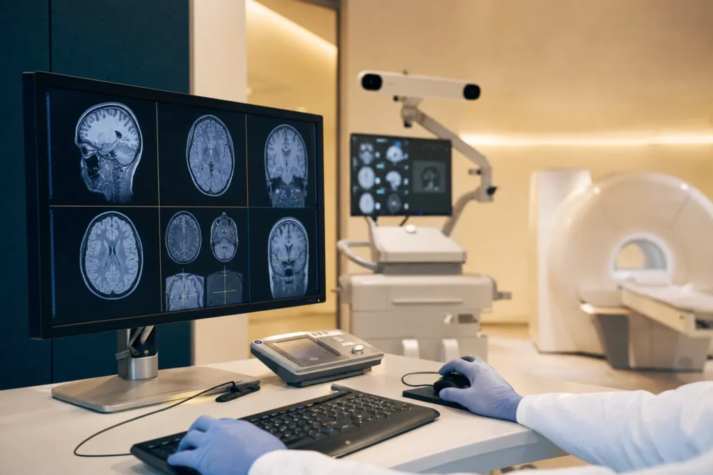
- MRI scan (magnetic resonance imaging) uses magnets and radio waves, with no radiation, to produce detailed pictures of the brain and spinal cord. It is often the most useful test. Young children may need a gentle sedative or a short general anaesthetic to keep still.
- CT scan (computed tomography) uses X-rays to give quick, detailed images and is especially useful in emergencies or for looking at the skull bones.
- Ultrasound can be used in babies whose soft spot is still open, to look at the fluid spaces in the brain without radiation.
Other tests may include blood tests, an EEG (a painless recording of the brain's electrical activity, used in epilepsy), or a lumbar puncture (taking a small sample of fluid from the lower back). If a tumour is suspected, a biopsy (taking a small piece of tissue to examine under a microscope) is often needed to find out exactly what type it is. For tumours, doctors also describe the type and how far it has spread; this information guides treatment and is explained by the team in terms relevant to your child.
Treatment options
Treatment is always tailored to the specific condition and child. Care is usually delivered by a multidisciplinary team (MDT) – a group that may include the neurosurgeon, paediatric anaesthetist, neurologist, oncologist (cancer specialist) if a tumour is present, radiologist, specialist nurses, physiotherapists, and others. They meet to agree the safest plan together.
Hydrocephalus. The two main approaches are a shunt — a thin, soft tube placed under the skin that drains excess fluid from the brain, usually into the abdomen (tummy), where the body absorbs it — and endoscopic third ventriculostomy (ETV), a keyhole operation that makes a small new opening so fluid can drain naturally without a permanent device. Shunts can sometimes block or become infected and may need adjusting over time.
Craniosynostosis. Surgery aims to relieve any pressure, give the brain room to grow, and improve head shape. This may be open surgery to remodel the skull, or a smaller endoscopic operation (sometimes followed by a moulding helmet). Timing is often in the first year of life.
Tumours. Treatment may combine surgery to remove as much of the tumour as is safe, with radiotherapy (carefully targeted high-energy beams) and/or chemotherapy (medicines that act on the tumour), depending on the type.
Spina bifida, Chiari malformation, tethered cord and epilepsy each have their own established surgical and supportive treatments, discussed in the sections that follow. Supportive care — pain relief, physiotherapy, and rehabilitation — is an important part of every plan.
Outlook: what to expect
Outlook varies enormously depending on the condition, so it is important to be guided by your child's own team rather than by general figures.
Many conditions can be treated effectively. Hydrocephalus that is treated promptly can often be well controlled, although some children need lifelong follow-up and occasional further surgery. Many children with craniosynostosis have no lasting problems after treatment, while some need ongoing support.
For childhood brain and spinal cord tumours, survival has improved over the years. According to the American Cancer Society, about 3 out of 4 children with brain tumours (all types combined) survive at least 5 years after diagnosis, but outcomes vary considerably by tumour type, location and other individual factors. It is essential to understand what these numbers are and are not. The American Cancer Society states plainly that survival rates "are only estimates" and "cannot tell you exactly what will happen with any one person." They are based on large groups of children treated in the past, and improvements in treatment since then may mean a better outlook today. These figures are population-level information, not a prediction for your child. Only your child's specialist, who knows the full picture, can discuss what is likely in your individual situation.
Living with it and follow-up
For many families, neurosurgery is the start of a longer journey of care rather than a single event. The shape of that journey depends on the condition.
Follow-up appointments and scans. Children are usually seen regularly so the team can check healing, watch growth and development, and pick up any problems early. A child with a shunt, for example, will be monitored over the years because shunts can occasionally block or become infected; parents are taught the warning signs to look out for.
Rehabilitation and development. Some children benefit from physiotherapy, occupational therapy, or speech and language therapy to support movement, daily skills and communication. Schools can often provide extra support so a child does not fall behind.
Emotional support. A diagnosis and surgery affect the whole family. It is normal for children and parents to feel anxious, and many hospitals offer psychological support, play specialists for younger children, and patient groups where families share experience. Looking after your own wellbeing helps you support your child.
Ask the team for a clear written plan: who to contact, which symptoms need urgent attention, and when the next review is due. Having this in plain language reduces uncertainty.
Planning treatment abroad: what affects cost and how to prepare records
If you are considering arranging your child's neurosurgery in another country, careful preparation makes everything smoother and safer. We do not list prices here, because the cost of paediatric neurosurgery is highly individual; the only reliable figure is a personalised estimate based on your child's actual records. The factors that most affect cost and planning include:
- the exact diagnosis and the type of operation needed (for example, a keyhole procedure versus open skull surgery);
- how many surgeries or treatments are planned, and whether radiotherapy or chemotherapy may also be involved;
- the type and length of imaging, anaesthesia and intensive monitoring required;
- the length of the hospital stay and any time in a paediatric intensive care unit;
- rehabilitation, follow-up scans and review appointments; and
- practical costs such as travel and accommodation for the child and family.
Preparing your records. Gather your child's recent MRI or CT scans (ideally the original image files, not only the reports), all written reports, a clear summary of the medical history, a list of current medicines and doses, and any biopsy or laboratory results. Having these ready allows a specialist to review the case properly and give an accurate, personalised estimate. A free consultation is the best way to find out what your child's specific situation involves before you commit to anything.
Why families consider Turkiye, and how to choose a good centre
Turkiye (Turkey) has become a well-known destination for medical care, with a number of large hospitals that treat international patients and offer paediatric services. Families often consider it for the combination of experienced specialists, modern facilities, and the convenience of coordinated international patient support. What matters most, though, is not the country but the quality and suitability of the specific centre for your child's condition.
When choosing a centre, it is reasonable to verify the following:
- Accreditation. Look for hospitals with recognised quality accreditation such as Joint Commission International (JCI), which assesses patient safety and care standards against international benchmarks.
- A genuine paediatric pathway. Check that there are paediatric neurosurgeons, paediatric anaesthetists, and a children's intensive care unit — children need teams trained specifically for their age group.
- Relevant experience. Ask how often the team treats your child's particular condition, and who exactly will be operating.
- A multidisciplinary team. For tumours especially, confirm that oncology, radiology and rehabilitation specialists are involved.
- Clear communication. Ensure you can get information, consent documents and a treatment plan in a language you understand, and that follow-up after you return home is discussed.
Seeking a second opinion is always reasonable and is something good doctors welcome. A trustworthy team will answer your questions openly and will never pressure you with promises of guaranteed results.
Prevention, second opinions and clinical trials
Most paediatric neurosurgical conditions cannot be prevented, and it is important for parents to know that these diagnoses are generally not caused by anything they did. There are, however, a few clear and helpful exceptions, along with sensible steps that support any child's recovery.
Folic acid before and during pregnancy significantly reduces the risk of neural tube defects such as spina bifida. Health services recommend that women take folic acid when planning a pregnancy and in early pregnancy. General safety measures — such as well-fitted helmets for cycling and appropriate car seats — reduce the risk of head injuries that can lead to neurosurgical problems.
Second opinions. For any major surgery, and especially for a tumour, you are entitled to seek a second opinion. This can give peace of mind and sometimes opens up additional options. Reputable specialists support this.
Clinical trials. For childhood tumours in particular, many children are treated as part of a clinical trial — a carefully supervised study comparing or refining treatments. Trials are how care continues to improve, and taking part is always voluntary and fully explained. Your child's team can tell you whether a relevant trial exists and whether it might be suitable. Whatever you decide, supportive care, good nutrition, follow-up and emotional support all help your child through treatment and recovery.
Frequently asked questions
What does a paediatric neurosurgeon do?
Is brain surgery on a child safe?
What is hydrocephalus and how is it treated?
What are the early signs of a brain problem in a baby?
Does craniosynostosis always need surgery?
How common are brain tumours in children?
What is the survival rate for childhood brain tumours?
Can paediatric neurosurgery conditions be prevented?
How is a diagnosis made?
What should I bring when seeking treatment abroad for my child?
How should I choose a hospital in Turkiye for my child?
Will my child need long-term follow-up?
Sources
- NHS — Hydrocephalus (overview, types, symptoms, treatment)
- Great Ormond Street Hospital (NHS) — Hydrocephalus
- NHS — Craniosynostosis
- NHS — Spina bifida
- NHS — Epilepsy treatment (including surgery)
- NINDS — Chiari malformation
- American Cancer Society — Brain and spinal cord tumours in children: key statistics
- American Cancer Society — Survival rates for childhood brain and spinal cord tumours
- Cancer Research UK — Children's brain and spinal cord tumours
- National Cancer Institute (cancer.gov) — Childhood brain and spinal cord tumours treatment (PDQ)
- Joint Commission International — Accreditation and accredited organizations
This article is for general information only and is not medical advice. Always consult a qualified doctor about your individual case.
Specialists in Paediatrics
Doctors of the department we work with directly. Which doctor takes your case is decided after the specialists have reviewed your records — your coordinator confirms it, not this page.



Considering this procedure?
Send us your photos and questions. A BergemHealth coordinator and a senior specialist will review your case and reply with honest, personalised guidance — no obligation.
Free consultation