Glioma (incl. glioblastoma)
If you or someone you love has just heard the word "glioma," it is natural to feel overwhelmed. This guide explains, in plain language, what a glioma is, how doctors tell the different types apart, what the symptoms and tests involve, and the treatments available today, including for glioblastoma. Our aim is to help you understand your situation clearly and calmly, so you can ask better questions and make informed decisions alongside a qualified specialist.
What a glioma is
A glioma is a tumour that begins in the glial cells of the brain or spinal cord. Glial cells are the brain's support cells: they surround the nerve cells (neurons) and help them work, feeding them, holding them in place, and keeping the chemistry around them balanced. When glial cells start to grow in an uncontrolled way, they can form a mass, which is what we call a tumour.
Because the brain and spinal cord sit inside the fixed space of the skull and spine, a growing glioma can press on nearby tissue. This pressure, rather than the tumour spreading to other organs, is usually what causes symptoms. Gliomas are described as primary brain tumours, meaning they start in the brain itself rather than spreading there from cancer somewhere else in the body.
Not all gliomas behave the same way. Some grow very slowly over years and are not considered cancerous in the everyday sense. Others, such as glioblastoma, grow quickly and invade healthy brain tissue. The type and grade of glioma matter a great deal, because they shape what treatment is recommended and what you can expect. Throughout this guide we explain each medical term as we go, so nothing is left as jargon.
Types and subtypes
Doctors classify gliomas in two ways at the same time: by the kind of glial cell the tumour resembles, and by how the cells look and behave under the microscope (its grade). Since 2016, and more extensively in the 2021 edition, the World Health Organisation (WHO) classification has also relied on molecular testing, looking at specific changes in the tumour's genes, because these markers predict behaviour better than appearance alone.
The main adult glioma types are:
- Astrocytoma develops from star-shaped glial cells called astrocytes. It ranges from slow-growing (low-grade) to aggressive (high-grade).
- Oligodendroglioma is a rarer glioma that starts in cells called oligodendrocytes. These tumours are graded 2 or 3 and tend to grow more slowly than glioblastoma.
- Ependymoma starts in cells lining the fluid-filled spaces of the brain and spinal cord. It is more common in children and is often lower-grade.
- Glioblastoma (sometimes still called glioblastoma multiforme, GBM) is the most aggressive glioma. It is always WHO grade 4 and is now considered a distinct diagnosis in its own right, not simply a high-grade astrocytoma. It is the most common malignant (cancerous) brain tumour in adults.
Two genetic markers shape the modern diagnosis. The IDH gene status matters most: an IDH-mutant tumour generally behaves less aggressively than an IDH-wildtype (unchanged) one. In oligodendroglioma, doctors also look for a change called 1p/19q co-deletion. These are not facts you need to memorise, but knowing the words helps you follow what your care team is saying.
Causes and risk factors
In honesty, doctors do not know what causes most gliomas. The tumour begins when glial cells develop changes (mutations) in their DNA, the set of instructions that tells a cell how to grow. These changes tell the cells to keep multiplying when they should stop, and to keep living when healthy cells would normally die. In the great majority of cases, these changes happen by chance during a person's lifetime, not because of anything they did or failed to do.
A few factors are linked to a higher risk, though they explain only a small share of cases:
- Age. Gliomas are most common in adults, with glioblastoma most often affecting people roughly between 45 and 70 years old. Some glioma types are more common in children.
- Previous radiation to the head. People who have had ionising radiation (for example, radiotherapy used to treat an earlier cancer) have a somewhat higher risk.
- Rare inherited conditions. A small number of genetic syndromes, such as neurofibromatosis, Li-Fraumeni syndrome and Turcot syndrome, raise the risk. Inherited gliomas are uncommon.
- Family history. Gliomas can occasionally run in families, but this is very rare and more research is needed.
It is worth saying plainly what does not have proven, accepted links: everyday mobile phone use is not established as a cause of glioma by the authorities cited here. If brain tumours run in your close biological family, you can ask about genetic counselling. For most people, though, a glioma is not something that could have been predicted or prevented.
Signs and symptoms (and when to see a doctor)
Glioma symptoms depend less on the tumour type and more on where it sits in the brain, how big it is, and how fast it is growing. A small tumour in a sensitive area can cause clear symptoms, while a larger one in a quieter region may cause few. Symptoms can build slowly over weeks and months, or, with faster-growing tumours, appear over a shorter time.
Common signs and symptoms include:
- Headaches, often worst in the morning, that are new or different from your usual headaches
- Nausea or vomiting
- Seizures (fits), especially a first-ever seizure in an adult
- Vision changes such as blurred or double vision, or loss of side vision
- Difficulty with speech, or trouble finding words
- Weakness or numbness, often on one side of the body
- Problems with balance or walking
- Changes in memory, thinking, mood or personality
When to see a doctor. Most headaches are not caused by a brain tumour, and most of the symptoms above have far more common, less serious explanations. Even so, you should arrange to see a doctor if you have a first-ever seizure, a headache that is new and persistent or steadily worsening, or any new weakness, vision loss, confusion or speech difficulty. Seek urgent help for a sudden severe headache unlike any before, a seizure that does not stop, or a rapid decline in alertness. Acting early simply means any problem can be looked into properly, not that the news will be bad.
Screening and early detection
There is currently no routine screening test for glioma in people without symptoms. Unlike breast or bowel cancer, brain tumours are not common enough in the general population, and there is no simple, safe test that reliably finds them early, so population-wide screening is not offered anywhere as standard. This is an honest limitation rather than an oversight.
In practice, gliomas are usually found when a person develops symptoms and has a brain scan, or sometimes by chance on a scan done for an unrelated reason (an incidental finding). For the small number of people with a known inherited syndrome that raises brain-tumour risk, specialists may recommend a personalised monitoring plan, which is different from general screening.
Because there is no screening programme, the most useful thing an ordinary person can do is take persistent new neurological symptoms seriously and get them checked. Early assessment does not change the underlying biology of a tumour, but it does mean treatment can be planned without unnecessary delay.
How it is diagnosed
Diagnosing a glioma usually happens in steps, and it is normal for this to take a little time. Your doctor will first ask about your symptoms and medical history and carry out a neurological examination, checking things like vision, balance, strength, reflexes, coordination and memory to see how the brain and nerves are working.
If a tumour is suspected, imaging follows:
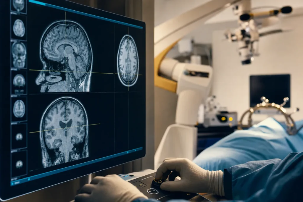
- MRI scan (magnetic resonance imaging) is the main test. It uses magnets and radio waves, not radiation, to produce detailed pictures of the brain. A contrast dye (gadolinium) is often given to make a tumour show up more clearly.
- CT scan may be used, especially if MRI is not possible, or in an urgent situation.
- Specialised scans such as PET scans or advanced MRI sequences are sometimes added to gather more detail.
Imaging can suggest a glioma, but a firm diagnosis almost always needs a biopsy, a small sample of tumour tissue examined under the microscope. This may be done as a separate procedure or as part of surgery to remove the tumour. A pathologist then assigns the grade (1 to 4) and carries out molecular testing for markers such as IDH and, in glioblastoma, the MGMT status, which can predict how well certain chemotherapy works. This combined tissue-and-molecular result is what gives the precise diagnosis that guides treatment. Gliomas are graded rather than "staged" in the way many other cancers are, because they rarely spread outside the brain and spinal cord.
Treatment options
Glioma treatment is planned by a multidisciplinary team, a group of specialists who meet to agree the best approach for each person. This typically includes a neurosurgeon, a neuro-oncologist (a doctor specialising in brain cancers), a radiation oncologist, a neuroradiologist, a pathologist, and specialist nurses, with input from rehabilitation and palliative care as needed. The plan depends on the tumour type, grade, molecular markers, its size and location, and your age and general health.
The main treatments are:
- Surgery. Often the first step when the tumour can be reached safely. The aim is maximal safe resection: removing as much tumour as possible without harming vital brain functions. The usual operation is a craniotomy (temporarily opening part of the skull). Sometimes part of the surgery is done while you are awake, so the team can check speech and movement as they work near important areas. For some tumours, minimally invasive laser ablation (using heat to destroy tumour tissue) may be an option.
- Radiation therapy (radiotherapy). High-energy beams target remaining tumour cells, usually after surgery. Modern techniques such as intensity-modulated radiotherapy (IMRT) and stereotactic radiosurgery focus the dose on the tumour while sparing healthy tissue.
- Chemotherapy. Medicines that stop cancer cells growing. For glioblastoma, the established approach combines radiotherapy with a drug called temozolomide, followed by further temozolomide afterwards, an approach widely known from the landmark Stupp trial. Drug-releasing wafers are sometimes placed in the cavity during surgery.
- Tumour treating fields (TTFields). A wearable device delivering low-intensity electric fields through pads on the scalp, used with maintenance temozolomide in some adults with glioblastoma to interfere with cancer-cell division.
- Targeted therapy and supportive medicines. For recurrent glioblastoma, a drug called bevacizumab may be used. Steroids reduce brain swelling, and anti-seizure medicines control fits.
For some slow-growing, low-grade gliomas, the team may recommend active surveillance, watching closely with regular scans and treating only if the tumour changes. There is no single right answer for everyone, which is exactly why a specialist team and a clear conversation matter.
Outlook: what to expect
Outlook varies enormously between glioma types, and this is one of the most important things to understand. A low-grade, IDH-mutant tumour in a younger adult is a very different situation from an IDH-wildtype glioblastoma. The figures below come from large cancer registries and describe 5-year relative survival, the percentage of people with that tumour still alive five years after diagnosis compared with the general population. They are population averages, not a prediction for any individual.
Drawing on US registry data (covering patients diagnosed 2004 to 2020) reported by the American Cancer Society, 5-year relative survival differs by tumour and by age:
- Oligodendroglioma: about 93% at ages 15 to 39, and about 79% at ages 40 and over.
- Low-grade diffuse astrocytoma: about 79% at ages 15 to 39, and about 34% at ages 40 and over.
- Anaplastic (grade 3) astrocytoma: about 64% at ages 15 to 39, and about 21% at ages 40 and over.
- Glioblastoma: about 28% at ages 15 to 39, and about 6% at ages 40 and over.
For glioblastoma specifically, sources describe a typical (median) survival of roughly 12 to 18 months with standard treatment, with a small proportion of people living considerably longer; results tend to be more favourable when the tumour has a methylated MGMT marker that responds well to temozolomide.
Please read these numbers gently. They reflect groups of people, including many treated years ago before today's approaches, and cannot tell you what will happen to you. Factors such as age, the molecular profile of the tumour, how much was removed at surgery, and general health all influence the picture. Your own specialist is the right person to discuss what these statistics mean in your particular case.
Living with it and follow-up
Living with a glioma is about more than the tumour itself; it is about daily life, energy, mood and relationships. After treatment you will have regular follow-up appointments and MRI scans to watch for any change or return of the tumour. These scans can bring anxiety (sometimes called "scanxiety"), and that is completely understandable; many centres can connect you with psychological support.
Recovery and rehabilitation often involve a wider team. Physiotherapy can help with strength and balance, occupational therapy with everyday tasks, and speech and language therapy if communication or swallowing is affected. Fatigue is one of the most common and underestimated effects of both the tumour and its treatment, and pacing your activity is a real and useful strategy.
Practical matters matter too. Seizures, certain medicines and the tumour's location can affect whether and when you are allowed to drive, so follow local rules and your team's advice. Many people are able to return to work, sometimes in an adjusted role. Lean on the support available: specialist nurses, brain-tumour charities, support groups and, importantly, your family and friends. You do not have to navigate this alone, and asking for help is a sign of good self-care, not weakness.
Planning treatment abroad: what affects cost and how to prepare your records
Some people choose to arrange glioma or glioblastoma treatment abroad, often to access an experienced neurosurgical team, modern equipment, or shorter waiting times. If you are considering this, it helps to understand the factors that influence cost so you can plan realistically. We do not publish prices here because every case is genuinely different; instead, the right next step is a personalised estimate based on your actual medical records.
The main factors that affect the cost and complexity of treatment include:
- The tumour's type, grade and molecular profile, which determine which treatments are needed
- The type and extent of surgery (for example, awake craniotomy or laser ablation), and the technology used
- Whether radiotherapy is needed, and how many sessions
- The chemotherapy regimen and its duration, and whether tumour treating fields are used
- Length of hospital stay and intensive-care needs
- Imaging, pathology and molecular testing
- Rehabilitation, follow-up scans, and any need for a longer stay
- Translation, accommodation and travel for you and a companion
To prepare, gather your medical records in advance: recent MRI and CT scans (ideally on a disc or in DICOM digital format, not just reports), the pathology and molecular-testing results, a summary of treatments so far, your current medicines, and a letter from your current doctor. Having these ready allows a specialist team to review your case properly and give a meaningful, individualised plan and estimate, rather than a generic figure. A free consultation is the simplest way to begin that conversation.
Why Turkiye, and how to choose a good centre
Turkiye (Turkey) has become a well-known destination for medical care, including neurosurgery, with a number of large hospitals that treat international patients and offer modern imaging, surgery and radiotherapy. For a serious condition like glioma, however, the destination matters less than the quality and experience of the specific team looking after you. The goal is the right care, not simply care abroad.
When choosing a centre, it is reasonable to verify the following:
- Accreditation. Look for recognised hospital accreditation, such as Joint Commission International (JCI), as a baseline marker of quality and safety standards.
- A genuine multidisciplinary team. Glioma care should involve neurosurgeons, neuro-oncologists, radiation oncologists, neuroradiologists and pathologists who discuss cases together, rather than a single doctor working alone.
- Relevant experience. Ask how often the team treats gliomas and glioblastoma specifically, and whether they offer modern tools such as awake surgery, intra-operative MRI or neuro-navigation where appropriate.
- Molecular pathology. Confirm the centre carries out the molecular testing (IDH, MGMT and others) that modern diagnosis and treatment rely on.
- Clear communication. Reliable interpreting, written treatment plans, and honest answers to your questions, including about risks, are signs of a trustworthy team.
- Continuity of care. Ask how follow-up, scans and any complications will be handled once you return home.
Be cautious of anyone who promises a cure, uses words like "best" or "guaranteed," or pressures you to decide quickly. A good centre will welcome a second opinion and give you the information and time you need.
Clinical trials and getting a second opinion
Because glioblastoma and high-grade gliomas remain difficult to treat, a great deal of research is underway, and clinical trials are a meaningful option for many people. Trials test new approaches, including targeted drugs, immunotherapies (treatments that enlist the body's own immune system), vaccines, and novel surgical and radiation techniques. Taking part is always voluntary, and your team can explain whether a suitable trial is open and what it would involve. Reputable trial information is available through national cancer bodies such as the National Cancer Institute.
A second opinion is also entirely reasonable and is something experienced specialists expect and respect. Brain-tumour diagnoses rely heavily on careful reading of scans and pathology, and another expert review can confirm the diagnosis, clarify the grade and molecular profile, and sometimes open up additional treatment options. Seeking one does not offend your current team and does not delay urgent care when arranged sensibly.
Finally, look after yourself alongside formal treatment. While there is no proven way to prevent glioma, general good health, staying as active as your condition allows, eating well, sleeping, and tending to your mental wellbeing all support you through treatment and recovery. Most of all, stay connected to a qualified specialist who knows your case, and let them help you weigh each decision. Clear information and steady support, more than any single statistic, are what help people face a glioma diagnosis.
Frequently asked questions
Is a glioma always cancer?
What is the difference between a glioma and a glioblastoma?
What are the first signs of a glioma?
How is a glioma diagnosed?
Can a glioma be cured?
What does the survival rate for glioblastoma mean for me?
Is there a screening test to catch glioma early?
What treatments are available for glioma and glioblastoma?
What affects the cost of glioma treatment abroad?
How do I choose a good centre for treatment in Turkiye?
Should I get a second opinion?
Can I prevent a glioma?
Sources
- Cleveland Clinic - Glioma: causes, symptoms, treatment and prognosis
- Cleveland Clinic - Glioblastoma (GBM)
- Mayo Clinic - Glioma: symptoms and causes
- National Cancer Institute - Adult Central Nervous System Tumors Treatment (PDQ), Patient Version
- Cancer Research UK - Glioma (brain tumours in adults)
- Cancer Research UK - Grades of brain and spinal cord tumours
- American Cancer Society - Survival rates for selected adult brain and spinal cord tumors
- NHS - Malignant brain tumour: treatment
- National Brain Tumor Society - Astrocytoma, IDH-mutant
This article is for general information only and is not medical advice. Always consult a qualified doctor about your individual case.
Specialists in Neurosurgery
Doctors of the department we work with directly. Which doctor takes your case is decided after the specialists have reviewed your records — your coordinator confirms it, not this page.




Considering this procedure?
Send us your photos and questions. A BergemHealth coordinator and a senior specialist will review your case and reply with honest, personalised guidance — no obligation.
Free consultation