Breast cancer
A breast cancer diagnosis raises many questions at once. This guide explains, in plain language and drawing on established cancer authorities, what breast cancer is, how it is found and staged, and the modern treatments available, so you and your medical team can make informed decisions together.
What breast cancer is
Breast cancer is a disease in which cells in the breast grow in an uncontrolled way and can form a tumour. Most breast cancers are carcinomas, and more specifically adenocarcinomas, meaning they begin in the gland-like cells that line the milk ducts (the tubes that carry milk to the nipple) or the lobules (the glands that make milk).
It is one of the most common cancers in women in many countries, including the United Kingdom and the United States. According to the American Cancer Society, an average woman in the US has roughly a 1 in 8 (about 13%) lifetime risk of developing breast cancer, and the median age at diagnosis is around 62. Breast cancer can also occur in men, though this is much less common.
It is important to keep this in perspective. According to the American Cancer Society, breast cancer death rates in the US have fallen substantially over recent decades, an overall decline attributed to earlier detection and better treatment. A diagnosis is serious, but outcomes are strongly influenced by the type of cancer and how early it is found, and these are matters to discuss with your medical team.
Types and subtypes
Breast cancer is described in two complementary ways: by where it starts and how far it has spread, and by the biological "receptors" on the cancer cells. Both descriptions shape treatment.
By location and invasion
- Ductal carcinoma in situ (DCIS) is a non-invasive (sometimes called pre-invasive) cancer. Abnormal cells are confined to the lining of a milk duct and have not spread into surrounding tissue.
- Invasive ductal carcinoma (IDC) begins in a duct and grows into nearby breast tissue. According to the American Cancer Society, it is the most common form, making up about 70 to 80% of breast cancers.
- Invasive lobular carcinoma (ILC) begins in the milk-producing lobules and spreads into nearby tissue.
- Less common types include inflammatory breast cancer (about 1 to 5% of cases, where cells block skin lymph vessels and the breast looks red and swollen), Paget disease of the nipple, and rarer tumours such as angiosarcoma and phyllodes tumours.
By receptor status (molecular subtype)
Pathology testing checks the tumour for three key markers, which help determine which systemic treatments are likely to help:
- Hormone-receptor positive (HR-positive): the cells carry receptors for oestrogen (ER) and/or progesterone (PR). These cancers, the most common group, can often be treated with hormone-blocking (endocrine) therapy.
- HER2-positive: the cells make too much of a protein called HER2, which can drive growth. These may respond to HER2-targeted drugs.
- Triple-negative breast cancer (TNBC): the cells lack ER, PR and excess HER2. According to the American Cancer Society, it accounts for about 15% of cases. It tends to grow and spread faster and does not respond to hormone or HER2 therapies, so chemotherapy and, in some cases, immunotherapy are central to treatment.
Risk factors and causes
In most cases there is no single cause, and breast cancer arises from a combination of factors. Having one or more risk factors does not mean you will develop the disease, and many people with breast cancer have no obvious risk factors at all.
Well-established factors recognised by cancer authorities include:
- Being a woman and getting older are among the strongest factors; risk rises with age.
- Inherited gene changes and family history, particularly faults in the BRCA1 and BRCA2 genes, and certain hereditary syndromes. These account for a minority of cases but can raise risk substantially.
- Reproductive and hormonal factors, such as starting periods early, a later menopause, having no children or a first child later in life, and use of combined menopausal hormone replacement therapy (HRT).
- Lifestyle and body-related factors, including drinking alcohol, being overweight or obese (especially after menopause), and physical inactivity.
- Dense breast tissue and previous radiotherapy to the chest, particularly at a young age.
Some of these factors, such as alcohol, weight and activity, can be modified and may lower risk, while others cannot. A doctor or genetic counsellor can help you understand your personal risk.
Signs and symptoms, and when to see a doctor
A well-known symptom is a new lump or thickening in the breast or armpit, which is often (but not always) painless. However, breast cancer can show in other ways, and not every change is caused by cancer.
Changes worth checking with a doctor include:
- A new lump or area of thickening in the breast or under the arm.
- A change in the size or shape of a breast.
- Skin changes, such as dimpling or puckering (sometimes likened to orange peel), redness, or flaky, peeling skin.
- Nipple changes, such as a nipple that turns inward, or a change in its appearance.
- Nipple discharge that is not breast milk, especially if it contains blood.
- Persistent pain in the breast or nipple area.
These symptoms are common with non-cancerous (benign) conditions too, so they are not proof of cancer. Even so, the advice from health services such as the NHS is consistent: if you notice a new or unusual change in your breast, see a doctor without delay so it can be checked. Finding breast cancer early can give a wider range of treatment options.
Screening and early detection
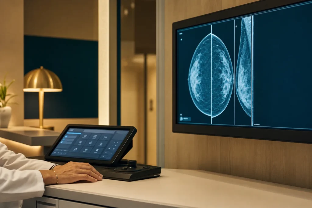
Screening means testing people who have no symptoms in order to find cancer early. For breast cancer the main screening tool is mammography, a low-dose X-ray of the breast, which can detect tumours before they can be felt.
Recommendations vary by country and by individual risk. As one example of an authoritative framework, the American Cancer Society advises women at average risk:
- Ages 40 to 44: the option to begin annual mammograms.
- Ages 45 to 54: a mammogram every year.
- Ages 55 and older: continue yearly, or switch to every two years, for as long as a woman is in good health with a life expectancy of at least 10 years.
National programmes (for example in the UK) use different age ranges and intervals, so follow the guidance where you live. Women at high risk, such as those with a known BRCA mutation, a strong family history, or previous chest radiation at a young age, may be advised to start earlier (the American Cancer Society suggests around age 30 for some groups) and to have breast MRI in addition to mammography. Being familiar with how your breasts normally look and feel, and reporting changes promptly, complements formal screening.
Diagnosis and staging
If a screening test or symptom raises concern, diagnosis usually follows a "triple assessment": a clinical examination, imaging (mammogram and/or ultrasound, sometimes MRI), and a biopsy in which a small tissue sample is taken and examined under a microscope. The biopsy confirms whether cancer is present and identifies the type and receptor status (ER, PR and HER2). Additional scans may be used to check whether the cancer has spread.
Staging describes how much cancer there is and where it is, which guides treatment and gives a sense of the outlook. Breast cancer staging combines several elements:
- The TNM system: T for the size of the tumour, N for whether nearby lymph nodes are involved, and M for whether it has spread (metastasised) to distant parts of the body.
- The grade, which describes how abnormal the cells look and how quickly they may grow.
- Biomarker status (ER, PR and HER2), and sometimes genomic tests that estimate recurrence risk.
These are combined into stages from 0 to IV. Stage 0 is non-invasive disease (DCIS). Stages I to III describe invasive cancer of increasing size and/or lymph node involvement that is still confined to the breast and nearby nodes. Stage IV means the cancer has spread to distant organs. Staging may be "clinical" (based on exams and scans) before treatment and "pathological" (based on tissue removed at surgery) afterwards.
Treatment options
Breast cancer treatment is highly individualised and often combines several approaches. Decisions are typically made by a multidisciplinary tumour board, a team that may include a breast surgeon, medical oncologist, radiation oncologist, radiologist, pathologist and specialist nurses. They weigh the cancer's stage and receptor status against your overall health and preferences.
Local treatments
- Surgery: the two main options are breast-conserving surgery (lumpectomy), which removes the tumour and a margin of healthy tissue while keeping most of the breast, and mastectomy, which removes the whole breast. For many early cancers, lumpectomy followed by radiotherapy can give results comparable to mastectomy; the right choice depends on tumour size and location, breast size, genetics and personal choice. Breast reconstruction may be offered.
- Lymph node surgery: a sentinel lymph node biopsy samples the first node(s) the cancer would reach, helping to stage the disease while sparing many people a more extensive node removal.
- Radiotherapy: targeted radiation, often used after breast-conserving surgery and sometimes after mastectomy, to lower the chance of the cancer returning.
Systemic (whole-body) treatments
- Chemotherapy: drugs that kill rapidly dividing cells, given before surgery (to shrink a tumour) or after (to reduce recurrence risk); central to triple-negative disease.
- Endocrine (hormone) therapy: for HR-positive cancers, medicines that block oestrogen's effect or lower oestrogen levels, usually taken for several years.
- HER2-targeted therapy: antibody-based and other targeted drugs for HER2-positive cancers.
- Immunotherapy: drugs that help the immune system attack cancer, used for some triple-negative cancers, often alongside chemotherapy.
Drug classes are described here in general terms; specific medicines and combinations are chosen by your oncology team.
Prognosis and survival
Survival statistics can help you understand the disease in general terms, but it is essential to read them carefully. They are population-level averages drawn from large groups of people diagnosed in the past. They are strongly stage-dependent, and they are not a prediction for any individual. An individual outlook depends on many factors, including the cancer's biology, age and health, and how the cancer responds to treatment, which is why your oncologist is the right person to discuss what the numbers mean for you.
To illustrate how much stage matters, the American Cancer Society reports the following 5-year relative survival rates for breast cancer in the US, based on women diagnosed between 2015 and 2021 (relative survival compares people with the cancer to the general population):
- Localised (confined to the breast): greater than 99%.
- Regional (spread to nearby structures or lymph nodes): about 87%.
- Distant (spread to organs such as the lungs, liver or bones): about 33%.
- All SEER stages combined: about 92%.
The American Cancer Society notes that because these figures come from people diagnosed years ago, they may not reflect the results of more recent treatments. They also do not capture individual circumstances, so use them only as broad context for a conversation with your care team.
Supportive and follow-up care
Good cancer care extends well beyond the main treatment. Supportive (palliative) care focuses on relieving symptoms and side effects, such as pain, fatigue, nausea or the effects of hormone therapy, and on emotional wellbeing. It can be provided at any stage, alongside treatment aimed at curing or controlling the cancer, and is not only for advanced disease.
After treatment, you will usually enter a programme of follow-up care. This typically includes regular check-ups, ongoing mammograms, and monitoring for side effects or signs that the cancer has returned. If you are taking long-term endocrine therapy, follow-up also supports staying on treatment and managing any side effects.
Many people benefit from rehabilitation and wellbeing support: physiotherapy (for example to manage shoulder movement or lymphoedema after lymph node surgery), help with bone health and menopausal symptoms, nutrition advice, psychological support, and peer or counselling services. Ask your team what survivorship support is available, and raise any new or persistent symptoms promptly.
Planning treatment abroad: what affects cost and how to prepare
If you are considering treatment in another country, careful planning makes the experience smoother and safer. Costs vary widely between countries and individuals, so rather than quoting figures, it helps to understand the factors that influence the overall cost of breast cancer care:
- The stage and subtype of the cancer, which influence how much treatment is needed.
- The type and extent of surgery (lumpectomy versus mastectomy, lymph node procedures, and any reconstruction).
- Whether radiotherapy is required, and how many sessions.
- The systemic therapies involved (chemotherapy, endocrine, HER2-targeted or immunotherapy) and their duration.
- Diagnostic and monitoring tests, including biopsies, scans and genomic tests.
- Length of hospital stay, plus accommodation, travel and interpreting if needed.
To prepare, gather your medical records in advance: pathology and biopsy reports (including ER, PR and HER2 results), imaging files and reports, a summary of treatments already received, and your medication list. Having these ready allows a centre to give accurate advice and a personalised estimate. The BergemHealth team can help you organise records and request a tailored plan through a consultation, so you can compare options with a clear picture of what is involved.
Turkiye and choosing a cancer centre
Turkiye (Turkey) has become a common destination for international patients seeking cancer care, with hospitals that offer modern surgery, radiotherapy and systemic treatments. As with any country, quality varies between centres, so the goal is to choose carefully rather than to assume any single hospital is right for everyone.
When evaluating a cancer centre, wherever it is located, it is reasonable to verify:
- A genuine multidisciplinary tumour board that reviews each case across surgery, medical oncology, radiation oncology, radiology and pathology.
- Accreditation and credentials, such as recognised hospital accreditation and the qualifications and experience of the breast surgeons and oncologists.
- Access to the full range of treatments you may need, including reconstruction, radiotherapy equipment, and modern systemic therapies, on one site or through clear referral pathways.
- Pathology and genomic testing capability, since accurate receptor and biomarker testing drives treatment.
- Clear communication, including written treatment plans, interpreting services, and continuity of care for follow-up once you return home.
A concierge service such as BergemHealth can help coordinate these checks, arrange a second opinion and manage logistics, while clinical decisions remain with the treating oncology team.
Clinical trials and second opinions
Clinical trials study new treatments or new ways of using existing ones. For some patients, a trial can offer access to investigational therapies under close monitoring, and trials are an option to discuss with your oncologist; suitability depends on the cancer type, stage and prior treatments.
Seeking a second opinion is a normal and accepted part of cancer care. A fresh review of your pathology, imaging and proposed plan can confirm the recommendation, surface alternative options, or simply give you confidence before proceeding. Many centres expect and welcome second opinions. If you are weighing treatment at home against treatment abroad, a second opinion is a sensible step, and BergemHealth can help arrange one as part of planning your care.
Frequently asked questions
Is a breast lump always cancer?
What are the main subtypes of breast cancer?
What does it mean if my cancer is triple-negative?
How are the stages of breast cancer determined?
When should I start breast cancer screening?
Is lumpectomy or mastectomy better?
Can the survival statistics tell me my personal outlook?
What should I prepare before seeking treatment abroad?
What affects the cost of breast cancer treatment?
Should I get a second opinion?
Can breast cancer affect men?
This article is for general information only and is not medical advice. Always consult a qualified doctor about your individual case.
Considering this procedure?
Send us your photos and questions. A BergemHealth coordinator and a department-head specialist will review your case and reply with honest, personalised guidance — no obligation.
Free consultation