Gastric & esophageal cancer
A diagnosis of stomach or esophageal cancer raises urgent, difficult questions. This guide explains, in plain language and drawing only on authoritative cancer sources, what these cancers are, how doctors diagnose and stage them, and the treatment paths available, so you and your family can make informed decisions, including whether to consider treatment abroad in Turkiye.
What gastric and esophageal cancer are
Gastric (stomach) cancer and esophageal cancer are distinct diseases, but they are often discussed together because the stomach and esophagus meet at one junction, they share several risk factors, and the cancers that form where they join behave similarly. Understanding the difference between them, and where they overlap, is the first step toward understanding treatment.
Stomach (gastric) cancer begins when cells in the lining of the stomach grow abnormally. According to the National Cancer Institute, most stomach cancers are adenocarcinomas, meaning they start in the mucus-producing cells of the innermost layer (the mucosa). The stomach wall is built in layers, from the mucosa on the inside through the submucosa, muscle layer, subserosa, and serosa on the outside. Cancer becomes more advanced as it grows from the inner lining into these deeper layers.
Esophageal cancer forms in the esophagus, the muscular tube that carries food from the throat to the stomach. The two main types are squamous cell carcinoma, which starts in the flat cells lining the esophagus (most often the upper and middle portions), and adenocarcinoma, which starts in glandular cells and usually forms in the lower esophagus near the stomach.
This guide covers both diseases. Throughout, remember that the information here is general and educational. Every person's situation is different, and decisions about diagnosis and treatment should always be made with a qualified oncologist and a multidisciplinary team.
Types and subtypes
Knowing the exact type and subtype matters because it shapes the whole treatment plan.
Gastric adenocarcinoma is classified in two useful ways. By location, the NCI distinguishes gastric cardia cancer, which begins in the top part of the stomach just below where it meets the esophagus, from non-cardia gastric cancer, which begins elsewhere in the stomach. By appearance under the microscope, pathologists describe an intestinal type (with cancer cells that look relatively similar to normal cells) and a diffuse type (with cells that look very different and tend to spread within the stomach wall).
Less common stomach tumors include gastrointestinal stromal tumors (GISTs), neuroendocrine tumors, and lymphomas; these are managed differently from adenocarcinoma and are outside the main scope of this guide.
Esophageal cancer has two principal subtypes:
- Adenocarcinoma typically arises in the lower esophagus and is linked to long-standing acid reflux and a related condition called Barrett's esophagus. It has become a more common type in many Western countries.
- Squamous cell carcinoma usually arises in the upper and middle esophagus and is more strongly linked to smoking and alcohol. It remains the most common esophageal cancer worldwide.
Cancers at the gastroesophageal (GE) junction, where the esophagus meets the stomach, sit between these categories and are often treated using approaches drawn from both gastric and esophageal protocols. Your care team will also test the tumor's biology, including HER2 status and certain immune markers, because these results can open up specific treatment options.
Risk factors and causes
Most risk factors raise the odds of developing cancer; they do not mean a person will certainly get it, and some people with no known risk factors still develop these diseases. The factors below are drawn from the NCI, the American Cancer Society, and Cancer Research UK.
Stomach cancer risk factors include:
- Helicobacter pylori (H. pylori) infection, a common stomach bacterium that raises the risk of cancer in the lower and middle stomach.
- Long-standing gastroesophageal reflux disease (GERD) and obesity, which may raise the risk of cancer in the upper stomach.
- A diet low in fruits and vegetables, or high in salted, smoked, or poorly preserved foods.
- Smoking tobacco.
- Chronic gastric inflammation, pernicious anemia, prior stomach surgery, certain inherited syndromes, and a family history of stomach cancer.
Esophageal cancer risk factors differ by subtype:
- For adenocarcinoma: Barrett's esophagus, long-term GERD, obesity, and smoking.
- For squamous cell carcinoma: smoking, heavy alcohol use, and a diet low in fruits and vegetables.
Barrett's esophagus deserves special mention. It is a condition in which the cells lining the lower esophagus are replaced by abnormal cells as a result of chronic acid exposure. It is not cancer, but it increases the risk of esophageal adenocarcinoma over time, which is why people with Barrett's are usually monitored. Treating H. pylori and managing reflux, along with not smoking, are among the steps that may lower risk; discuss prevention with your doctor.
Signs and symptoms, and when to see a doctor
Early gastric and esophageal cancers often cause few or no symptoms, and when symptoms do appear they overlap with far more common, non-cancerous conditions. That overlap is exactly why persistent or unusual symptoms should be checked rather than ignored.
Possible symptoms of stomach cancer (per the NHS and NCI) include indigestion or heartburn that does not go away, feeling full quickly when eating, difficulty swallowing, nausea or vomiting, pain in the upper abdomen, unintentional weight loss, tiredness, or a lump in the upper abdomen.
Possible symptoms of esophageal cancer (per Cancer Research UK) center on difficulty swallowing (dysphagia), sometimes described as food sticking in the throat or chest, or pain on swallowing. Other signs include persistent indigestion or heartburn, unexplained weight loss, chest or throat pain, food coming back up, a persistent cough, hoarseness, or, less commonly, blood in vomit or dark stools.
When to see a doctor: The NHS advises seeing a GP if you have difficulty swallowing, a lump, noticeable weight loss, or digestive symptoms that get worse or do not improve after three weeks. As the NHS emphasizes, these symptoms are very common and usually have other causes, but early evaluation matters because it gives a better chance of finding disease at a treatable stage.
Screening and early detection
Screening means testing people who have no symptoms in order to find cancer early. The picture differs for these two cancers and by region.
Stomach cancer: The NCI states that in the United States there are no standard or routine screening tests for people at average risk. However, upper endoscopy may be considered for people at higher risk, such as those with chronic gastric atrophy or pernicious anemia, a prior partial gastrectomy, certain inherited syndromes, a strong family history, or origins from regions where stomach cancer is more common. In some high-incidence countries, organized endoscopic screening programs exist; whether screening is appropriate for you is a question for your doctor.
Esophageal cancer: There is likewise no routine population screening. For people with Barrett's esophagus, doctors often recommend periodic endoscopic surveillance with biopsies to look for precancerous changes, so they can be treated before cancer develops.
If you have ongoing reflux, a family history, or other risk factors, ask your doctor whether endoscopic evaluation or surveillance makes sense for your situation. Acting on persistent symptoms remains the most important route to early detection for most people.
Diagnosis and staging
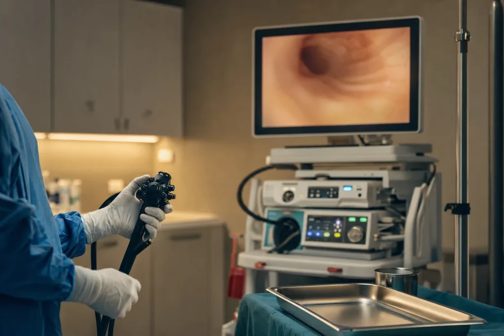
Diagnosis usually begins with upper endoscopy (gastroscopy), in which a thin, flexible tube with a camera is passed into the esophagus and stomach so the doctor can see the lining and take biopsies. A pathologist examines the tissue to confirm cancer, identify the type and subtype, and run molecular tests such as HER2 status and immune markers (for example PD-L1 expression and mismatch-repair/microsatellite-instability status), which can guide drug treatment.
Once cancer is confirmed, staging determines how far it has spread. Tests may include CT scans of the chest, abdomen, and pelvis; endoscopic ultrasound (EUS) to judge how deeply the tumor has grown into the wall and whether nearby lymph nodes are involved; PET-CT in selected cases; and sometimes diagnostic laparoscopy for stomach cancer to check the abdominal lining.
Both cancers use the TNM system (Tumor, Nodes, Metastasis) to assign a stage. For stomach cancer, the NCI describes the stages this way in simplified terms:
- Stage 0: abnormal cells confined to the mucosa (carcinoma in situ).
- Stage I: cancer in the inner layers, with limited or no lymph node involvement.
- Stage II: deeper growth into the wall and/or more lymph nodes involved.
- Stage III: extensive growth through the wall layers, more lymph nodes, and possible spread to nearby organs.
- Stage IV: cancer has spread to distant parts of the body, such as the liver, lungs, or distant lymph nodes.
Esophageal cancer is staged on similar TNM principles. Stage directly shapes whether treatment aims to cure the disease or to control it and relieve symptoms, so accurate staging is essential before any plan is finalized.
Treatment options
Treatment depends on the cancer type, its location, the stage, the tumor's molecular features, and your overall health. Most people receive a combination of treatments, planned and coordinated by a multidisciplinary team. The summaries below reflect the American Cancer Society and NCI; the right combination for any individual must be decided by their own oncology team.
Surgery. For stomach cancer, surgery may involve subtotal (partial) gastrectomy or total gastrectomy, removing part or all of the stomach together with nearby lymph nodes. For esophageal cancer, esophagectomy removes part of the esophagus and reconstructs the digestive tract. Very early, superficial tumors in either organ may sometimes be removed through the endoscope using endoscopic mucosal resection (EMR) or endoscopic submucosal dissection (ESD).
Chemotherapy. Drug treatment is a cornerstone. It is frequently given perioperatively, meaning before and after surgery, to shrink the tumor and reduce the chance of recurrence, and it is also used for advanced disease.
Radiation therapy and chemoradiation. Radiation is often combined with chemotherapy (chemoradiation), particularly before surgery for esophageal cancer or for locally advanced disease that cannot be removed surgically.
Targeted therapy. When a tumor overexpresses the HER2 protein, HER2-directed antibody therapy may be added to chemotherapy. Other targeted options exist for specific molecular changes; these are matched to test results.
Immunotherapy. Drugs that block the PD-1/PD-L1 pathway may be used, sometimes with chemotherapy, especially when the tumor expresses PD-L1 or shows high microsatellite instability (MSI-H) or a mismatch-repair defect (dMMR).
The multidisciplinary tumour board. Because these cancers are complex, decisions are best made in a tumour board: a meeting of surgical oncologists, medical oncologists, radiation oncologists, gastroenterologists, radiologists, and pathologists who review your case together and agree on a coordinated plan. This team approach is central to modern care at quality centres worldwide.
Prognosis and survival
Survival statistics describe what has happened, on average, to large groups of people diagnosed in the past. They are not a prediction for any individual. As the SEER program of the NCI cautions, because these figures are based on large groups, they cannot tell you exactly what will happen in your case, and outcomes depend on many factors, including stage, tumor biology, overall health, treatment, and how an individual responds. Treatments also continue to improve over time, which past statistics may not fully capture.
With those important caveats, the NCI's SEER data (2016 to 2022) report 5-year relative survival as follows.
Stomach cancer:
- Localized (confined to the stomach): about 78%
- Regional (spread to nearby structures or lymph nodes): about 39%
- Distant (spread to distant organs): about 8%
- All stages combined: about 40%
Esophageal cancer:
- Localized: about 49%
- Regional: about 29%
- Distant: about 5%
- All stages combined: about 22%
The clear pattern across both diseases is that outcomes are generally better when cancer is found and treated earlier. These numbers can help frame a conversation with your oncologist, who can place them in the context of your specific diagnosis, but they should never be read as a personal forecast.
Supportive and follow-up care
Good cancer care extends well beyond treating the tumor itself. Supportive (palliative) care focuses on relieving symptoms and improving quality of life, and it can be provided alongside treatment aimed at controlling or curing the cancer, at any stage.
Because both cancers can affect eating and swallowing, nutritional support is often important. A dietitian may help manage weight loss, and procedures such as placing a stent to keep the esophagus open, or a feeding tube, can help when swallowing is difficult. Pain management, anti-nausea treatment, and emotional and psychological support for the patient and family are all part of comprehensive care.
After treatment, follow-up care typically includes regular check-ups, scans, blood tests, and sometimes endoscopy to watch for recurrence and to manage longer-term effects of surgery, such as changes in digestion and how the body absorbs nutrients after part or all of the stomach or esophagus has been removed. Your team will set a follow-up schedule tailored to your treatment and recovery.
Planning treatment abroad: what affects cost and how to prepare records
Some patients and families consider treatment in another country, often to access experienced high-volume centres or shorter waiting times. If you are weighing this option, it helps to understand the factors that influence the overall cost of care, rather than any single price, because every treatment plan is different.
Cost is shaped by:
- The cancer type, subtype, and stage, and therefore the combination of treatments needed (surgery, chemotherapy, radiation, targeted or immunotherapy).
- The specific operation (for example, total versus subtotal gastrectomy, or esophagectomy) and whether minimally invasive techniques are used.
- The number of chemotherapy or radiation cycles and which drug classes are required, including whether HER2-directed or immunotherapy agents are part of the plan.
- Length of hospital stay, intensive care needs, and management of any complications.
- Diagnostic and staging tests, pathology and molecular testing, and follow-up.
- Accommodation, travel, interpretation, and length of stay for the patient and any companion.
To prepare, gather a complete medical record: pathology and biopsy reports, slides or blocks if available for review, imaging (CT, EUS, PET-CT) on disc, endoscopy reports, a list of current medications, and a summary of your medical history. Having these organized allows a centre to review your case accurately and give you a personalised plan. For an individual estimate based on your records, request a consultation; figures cannot be given meaningfully without reviewing your specific situation.
Why Turkiye, and how to choose a cancer centre
Turkiye (Turkey) has become a well-known destination for international patients seeking treatment, including for gastrointestinal cancers. The most important consideration is not the country but the quality and suitability of the specific centre and team caring for you. Choosing well matters far more than choosing fast.
When evaluating any cancer centre, in Turkiye or anywhere, it is reasonable to verify:
- A genuine multidisciplinary team and tumour board. Confirm that surgical, medical, and radiation oncologists, along with gastroenterology, radiology, and pathology, review cases together. This collaborative model underpins quality care.
- Accreditation. Look for recognized hospital accreditation and quality standards, and ask how the centre measures and reports its outcomes.
- Experience and volume. Centres that regularly perform gastrectomy and esophagectomy, and that manage these cancers frequently, tend to have well-developed pathways for complex care.
- Comprehensive services. Confirm access to molecular and HER2 testing, the full range of treatments (surgery, radiation, systemic therapy), and supportive and nutritional care.
- Clear communication. Reliable interpretation, transparent treatment plans, and a single point of contact help you make informed decisions throughout.
A medical-tourism concierge such as BergemHealth can help coordinate records, second opinions, and logistics, but the clinical plan should always rest with qualified oncologists. Take the time to ask questions and confirm these points before committing to care.
Clinical trials and second opinions
Clinical trials study new treatments and new combinations of existing treatments. For some patients they offer access to approaches not yet widely available, and they are an important part of how cancer care improves. Eligibility depends on factors such as cancer type, stage, prior treatment, and overall health. The NCI maintains information on trials, and your oncologist can advise whether a trial is appropriate and available for your situation.
A second opinion is a normal and reasonable step, especially before major surgery or when treatment options are complex or unclear. Another experienced specialist or tumour board may confirm the plan or suggest alternatives, and most clinicians welcome this. Seeking a second opinion does not usually cause harmful delay, and it can give you greater confidence in the path you choose. Whatever you decide, ensure that decisions are made together with a qualified oncology team that knows your full history.
Frequently asked questions
What is the difference between gastric and esophageal cancer?
What are the early symptoms of gastric cancer?
What is the main symptom of esophageal cancer?
What are the stages of gastric cancer?
Does H. pylori cause stomach cancer?
Is there screening for stomach or esophageal cancer?
What treatments are used for these cancers?
What is the survival rate for gastric and esophageal cancer?
What is HER2 testing and why does it matter?
What records should I prepare before seeking treatment abroad?
Should I get a second opinion?
Sources
- National Cancer Institute - What Is Stomach Cancer?
- National Cancer Institute - Stomach Cancer Causes and Risk Factors
- National Cancer Institute - Stages of Stomach Cancer
- National Cancer Institute - Stomach Cancer Screening
- National Cancer Institute - Esophageal Cancer Treatment (PDQ)
- American Cancer Society - Treating Stomach Cancer
- American Cancer Society - Treating Esophagus Cancer
- American Cancer Society - Risk Factors for Esophageal Cancer
- NCI SEER - Cancer Stat Facts: Stomach Cancer
- NCI SEER - Cancer Stat Facts: Esophageal Cancer
- NHS - Stomach Cancer Symptoms
- Cancer Research UK - Oesophageal Cancer Symptoms
This article is for general information only and is not medical advice. Always consult a qualified doctor about your individual case.
Specialists in Oncology
Doctors of the department we work with directly. Which doctor takes your case is decided after the specialists have reviewed your records — your coordinator confirms it, not this page.



Considering this procedure?
Send us your photos and questions. A BergemHealth coordinator and a senior specialist will review your case and reply with honest, personalised guidance — no obligation.
Free consultation